Advertisement

Main menu

  • Home
  • Content
    • Current Issue
    • Accepted Manuscripts
    • Article Preview
    • Past Issue Archive
    • Video Articles
    • AJNR Case Collection
    • Case of the Week Archive
    • Case of the Month Archive
    • Classic Case Archive
  • Special Collections
    • AJNR Awards
    • ASNR Foundation Special Collection
    • Most Impactful AJNR Articles
    • Photon-Counting CT
    • Spinal CSF Leak Articles (Jan 2020-June 2024)
  • Multimedia
    • AJNR Podcasts
    • AJNR SCANtastic
    • Trainee Corner
    • MRI Safety Corner
    • Imaging Protocols
  • For Authors
    • Submit a Manuscript
    • Submit a Video Article
    • Submit an eLetter to the Editor/Response
    • Manuscript Submission Guidelines
    • Statistical Tips
    • Fast Publishing of Accepted Manuscripts
    • Graphical Abstract Preparation
    • Imaging Protocol Submission
    • Author Policies
  • About Us
    • About AJNR
    • Editorial Board
    • Editorial Board Alumni
  • More
    • Become a Reviewer/Academy of Reviewers
    • Subscribers
    • Permissions
    • Alerts
    • Feedback
    • Advertisers
    • ASNR Home

User menu

  • Alerts
  • Log in

Search

  • Advanced search
American Journal of Neuroradiology

American Journal of Neuroradiology

ASHNR American Society of Functional Neuroradiology ASHNR American Society of Pediatric Neuroradiology ASSR
  • Alerts
  • Log in

Advanced Search

  • Home
  • Content
    • Current Issue
    • Accepted Manuscripts
    • Article Preview
    • Past Issue Archive
    • Video Articles
    • AJNR Case Collection
    • Case of the Week Archive
    • Case of the Month Archive
    • Classic Case Archive
  • Special Collections
    • AJNR Awards
    • ASNR Foundation Special Collection
    • Most Impactful AJNR Articles
    • Photon-Counting CT
    • Spinal CSF Leak Articles (Jan 2020-June 2024)
  • Multimedia
    • AJNR Podcasts
    • AJNR SCANtastic
    • Trainee Corner
    • MRI Safety Corner
    • Imaging Protocols
  • For Authors
    • Submit a Manuscript
    • Submit a Video Article
    • Submit an eLetter to the Editor/Response
    • Manuscript Submission Guidelines
    • Statistical Tips
    • Fast Publishing of Accepted Manuscripts
    • Graphical Abstract Preparation
    • Imaging Protocol Submission
    • Author Policies
  • About Us
    • About AJNR
    • Editorial Board
    • Editorial Board Alumni
  • More
    • Become a Reviewer/Academy of Reviewers
    • Subscribers
    • Permissions
    • Alerts
    • Feedback
    • Advertisers
    • ASNR Home
  • Follow AJNR on Twitter
  • Visit AJNR on Facebook
  • Follow AJNR on Instagram
  • Join AJNR on LinkedIn
  • RSS Feeds

AJNR is seeking candidates for the AJNR Podcast Editor. Read the position description.

Research ArticleADULT BRAIN
Open Access

Cortical Perfusion Alteration in Normal-Appearing Gray Matter Is Most Sensitive to Disease Progression in Relapsing-Remitting Multiple Sclerosis

S.-P. Hojjat, M. Kincal, R. Vitorino, C.G. Cantrell, A. Feinstein, L. Zhang, L. Lee, P. O'Connor, T.J. Carroll and R.I. Aviv
American Journal of Neuroradiology August 2016, 37 (8) 1454-1461; DOI: https://doi.org/10.3174/ajnr.A4737
S.-P. Hojjat
cMedical Imaging (S.-P.H., M.K., R.V., R.I.A., L.Z.), Sunnybrook Health Sciences Centre, Toronto, Ontario, Canada
fMedical Imaging (S.-P.H., R.I.A.), University of Toronto, Toronto, Ontario, Canada
  • Find this author on Google Scholar
  • Find this author on PubMed
  • Search for this author on this site
  • ORCID record for S.-P. Hojjat
M. Kincal
cMedical Imaging (S.-P.H., M.K., R.V., R.I.A., L.Z.), Sunnybrook Health Sciences Centre, Toronto, Ontario, Canada
  • Find this author on Google Scholar
  • Find this author on PubMed
  • Search for this author on this site
  • ORCID record for M. Kincal
R. Vitorino
cMedical Imaging (S.-P.H., M.K., R.V., R.I.A., L.Z.), Sunnybrook Health Sciences Centre, Toronto, Ontario, Canada
  • Find this author on Google Scholar
  • Find this author on PubMed
  • Search for this author on this site
  • ORCID record for R. Vitorino
C.G. Cantrell
gDepartments of Biomedical Engineering (C.G.C., T.J.C.)
  • Find this author on Google Scholar
  • Find this author on PubMed
  • Search for this author on this site
  • ORCID record for C.G. Cantrell
A. Feinstein
aFrom the Departments of Psychiatry (A.F.)
ePsychiatry (A.F.)
  • Find this author on Google Scholar
  • Find this author on PubMed
  • Search for this author on this site
  • ORCID record for A. Feinstein
L. Zhang
cMedical Imaging (S.-P.H., M.K., R.V., R.I.A., L.Z.), Sunnybrook Health Sciences Centre, Toronto, Ontario, Canada
  • Find this author on Google Scholar
  • Find this author on PubMed
  • Search for this author on this site
  • ORCID record for L. Zhang
L. Lee
bNeurology (L.L.)
dDepartments of Medicine (P.O., L.L.)
  • Find this author on Google Scholar
  • Find this author on PubMed
  • Search for this author on this site
  • ORCID record for L. Lee
P. O'Connor
dDepartments of Medicine (P.O., L.L.)
  • Find this author on Google Scholar
  • Find this author on PubMed
  • Search for this author on this site
  • ORCID record for P. O'Connor
T.J. Carroll
gDepartments of Biomedical Engineering (C.G.C., T.J.C.)
hRadiology (T.J.C.), Northwestern University, Chicago, Illinois.
  • Find this author on Google Scholar
  • Find this author on PubMed
  • Search for this author on this site
  • ORCID record for T.J. Carroll
R.I. Aviv
cMedical Imaging (S.-P.H., M.K., R.V., R.I.A., L.Z.), Sunnybrook Health Sciences Centre, Toronto, Ontario, Canada
fMedical Imaging (S.-P.H., R.I.A.), University of Toronto, Toronto, Ontario, Canada
  • Find this author on Google Scholar
  • Find this author on PubMed
  • Search for this author on this site
  • ORCID record for R.I. Aviv
  • Article
  • Figures & Data
  • Supplemental
  • Info & Metrics
  • Responses
  • References
  • PDF
Loading

Article Figures & Data

Figures

  • Tables
  • Fig 1.
    • Download figure
    • Open in new tab
    • Download powerpoint
    Fig 1.

    Block diagram representation of the image acquisition and processing pipeline. PSIR indicates phase-sensitive inversion recovery; PD, proton density.

  • Fig 2.
    • Download figure
    • Open in new tab
    • Download powerpoint
    Fig 2.

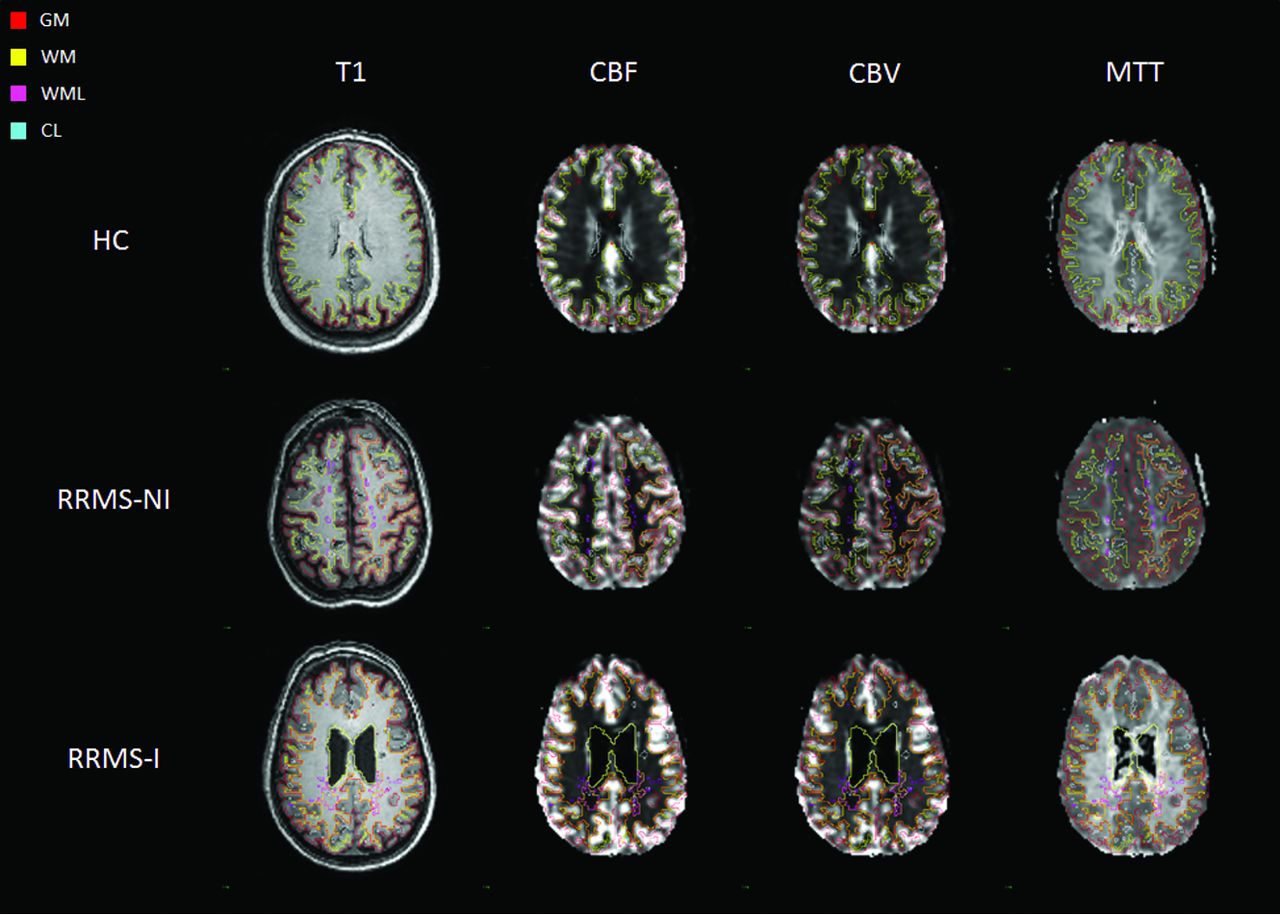
    Representative section of T1 and perfusion images in perfusion space for the different study groups with overlays of the investigated ROIs.

  • Fig 3.
    • Download figure
    • Open in new tab
    • Download powerpoint
    Fig 3.

    Distribution of the absolute perfusion cortical lesion results in the construction of 4 different populations: Standard CL (neither CBF nor CBV outliers), CBF outliers, CBV outliers, and CBV-CBF outliers (CBV outliers that are also CBF outliers). Absolute values of cerebral blood flow and cerebral blood volume are reported on the x-axis and y-axis, respectively. Each RRMS-NI lesion (A) is represented by a circle, and RRMS-I lesion (B) is represented by a triangle.

  • Fig 4.
    • Download figure
    • Open in new tab
    • Download powerpoint
    Fig 4.

    Distribution of the absolute perfusion white matter lesion results in the construction of 4 different populations by using the interquartile range method: Standard WML (neither CBF nor CBV outliers), CBF outliers, CBV outliers, and CBV-CBF outliers (CBV outliers that are also CBF outliers). Absolute values of cerebral blood flow and cerebral blood volume are reported on the x-axis and y-axis, respectively. Each RRMS-NI lesion (A) is represented by circle, and RRMS-I lesion (B) is represented by a triangle.

Tables

  • Figures
    • View popup
    Table 1:

    Participant demographic data, neurocognitive scores, and fractional brain volumea

    HC (n = 19)RRMS-NI (n = 19)RRMS-I (n = 20)
    Demographics
        Age (yr)49.0 ± 7.146.4 ± 7.248.1 ± 4.7
        Female sex (No.) (%)14 (73.68)15 (78.95)12 (60)
        Education (yr)16.9 ± 2.9b16.1 ± 1.314.6 ± 1.9b
        Disease duration (yr)0.0 ± 0.011.8 ± 5.411.6 ± 4.9
        HADS-A (log) (median) (IQR)3 (1–6)b,c6 (5–7)c8 (7–10)b
        HADS-D (log) (median) (IQR)2 (1–3)b3 (1,5)d8 (6–10)b,d
        EDSS median (IQR)NA1.5 (1–2)d2.5 (2–3)d
    Percentage fractional brain volume
        BPF79 ± 9b75 ± 672 ± 8b
        fC45.15 ± 5.1243.43 ± 3.9141.87 ± 5.50
        fWM31.66 ± 4.07b29.61 ± 2.8328.35 ± 3.5b
        fCL0.00 ± 0.00b0.01 ± 0.010.01 ± 0.02b
        fBG1.35 ± 0.191.31 ± 0.181.25 ± 0.22
        fTh0.68 ± 0.12b0.64 ± 0.140.55 ± 0.14b
        fWML0.00 ± 0.00b0.67 ± 0.740.92 ± 0.90b
        fT1 hole0.00 ± 0.00b0.23 ± 0.220.410 ± 0.506b
        fCSF21.16 ± 8.78b24.10 ± 6.2426.64 ± 7.32b
    • Note:—HADS indicates Hospital Anxiety [A] and Depression [D] Scale; EDSS, Extended Disability Status Scale; BPF, brain parenchymal fraction; fC, fractional cortical volume; fWM, fractional white matter volume; fCL, fractional cortical lesions volume; fBG, fractional basal ganglia volume; fTh, fractional thalamus volume; fWML, fractional white matter lesions volume; fTI hole, fractional T1 hole volume; fCSF, fractional CSF volume; HC, healthy controls; NA, not applicable; IQR, interquartile range.

    • ↵a All values are means unless otherwise specified. Significant P value < .017.

    • ↵b HC vs RRMS-I.

    • ↵c HC vs RRMS-NI.

    • ↵d RRMS-NI vs RRMS-I.

    • View popup
    Table 2:

    Comparison of perfusion parameters between study groupsa

    HC (n = 19)RRMS-NI (n = 19)RRMS-I (n = 20)
    WMGMWMLNAWMCLNAGMWMLNAWMCLNAGM
    CBF21.8b44.0f14.4d,e21.5d,e21.4c41.1c,d,f12.5d,e17.0b,d,e23.1c31.7c,d,f
    (mL/100 g per min)(19.7–28.4)(37.9–49.5)(9.9–20.2)(16.0–29.5)(15.7–32.3)(29.8–55.7)(8.1–18.5)(13.2–25.2)(15.4–34.3)(24.6–44.5)
    CBV1.5b2.8f1.1d,e1.6d,e1.4c2.5c,d1.0d,e1.3b,d.e1.7c2.1c,d,f
    (mL/100 g)(1.2–1.9)(2.1–3.2)(0.8–1.6)(1.2–2.1)(1.1–2.0)(1.9–3.4)(0.7–1.4)(1.0–1.7)(1.2–2.4)(1.7–2.7)
    MTT4.3b3.8f4.8e4.6e4.33.9d,f5.0e4.8b,e4.6c4.0c,d,f
    (min)(3.9–5.0)(3.4–4.3)(4.1–5.9)(4.1–5.2)(3.5–4.9)(3.3–4.4)(4.2–6.0)(4.3–5.2)(3.9–5.4)(3.5–4.6)
    • Note:—HC indicates healthy controls.

    • ↵a Normal-appearing GM and WM tissues are compared across patients with RRMS who were cognitively unimpaired and impaired and with healthy controls with corrected P < .017 (ie, .05/3) identified as significantly different. Lesion perfusion was also independently compared across disease groups and with the corresponding normal-appearing tissue within each patient group with P < .05 identified as significantly different. The values represent medians; the interquartile range for values are giving in parentheses.

    • ↵b HC WM vs NAWM.

    • ↵c CL vs NAGM.

    • ↵d RRMS-NI vs RRMS-I.

    • ↵e WML vs NAWM.

    • ↵f HC GM vs NAGM.

PreviousNext
Back to top

In this issue

American Journal of Neuroradiology: 37 (8)
American Journal of Neuroradiology
Vol. 37, Issue 8
1 Aug 2016
  • Table of Contents
  • Index by author
  • Complete Issue (PDF)
Advertisement
Print
Download PDF
Email Article

Thank you for your interest in spreading the word on American Journal of Neuroradiology.

NOTE: We only request your email address so that the person you are recommending the page to knows that you wanted them to see it, and that it is not junk mail. We do not capture any email address.

Enter multiple addresses on separate lines or separate them with commas.
Cortical Perfusion Alteration in Normal-Appearing Gray Matter Is Most Sensitive to Disease Progression in Relapsing-Remitting Multiple Sclerosis
(Your Name) has sent you a message from American Journal of Neuroradiology
(Your Name) thought you would like to see the American Journal of Neuroradiology web site.
CAPTCHA
This question is for testing whether or not you are a human visitor and to prevent automated spam submissions.
Cite this article
S.-P. Hojjat, M. Kincal, R. Vitorino, C.G. Cantrell, A. Feinstein, L. Zhang, L. Lee, P. O'Connor, T.J. Carroll, R.I. Aviv
Cortical Perfusion Alteration in Normal-Appearing Gray Matter Is Most Sensitive to Disease Progression in Relapsing-Remitting Multiple Sclerosis
American Journal of Neuroradiology Aug 2016, 37 (8) 1454-1461; DOI: 10.3174/ajnr.A4737

Citation Manager Formats

  • BibTeX
  • Bookends
  • EasyBib
  • EndNote (tagged)
  • EndNote 8 (xml)
  • Medlars
  • Mendeley
  • Papers
  • RefWorks Tagged
  • Ref Manager
  • RIS
  • Zotero
0 Responses
Respond to this article
Share
Bookmark this article
Cortical Perfusion Alteration in Normal-Appearing Gray Matter Is Most Sensitive to Disease Progression in Relapsing-Remitting Multiple Sclerosis
S.-P. Hojjat, M. Kincal, R. Vitorino, C.G. Cantrell, A. Feinstein, L. Zhang, L. Lee, P. O'Connor, T.J. Carroll, R.I. Aviv
American Journal of Neuroradiology Aug 2016, 37 (8) 1454-1461; DOI: 10.3174/ajnr.A4737
del.icio.us logo Twitter logo Facebook logo Mendeley logo
  • Tweet Widget
  • Facebook Like
  • Google Plus One
Purchase

Jump to section

  • Article
    • Abstract
    • ABBREVIATIONS:
    • Materials and Methods
    • Results
    • Discussion
    • Conclusions
    • Footnotes
    • References
  • Figures & Data
  • Supplemental
  • Info & Metrics
  • Responses
  • References
  • PDF

Related Articles

  • PubMed
  • Google Scholar

Cited By...

  • No citing articles found.
  • Crossref (20)
  • Google Scholar

This article has been cited by the following articles in journals that are participating in Crossref Cited-by Linking.

  • The role of brain vasculature in neurodegenerative disorders
    Melanie D. Sweeney, Kassandra Kisler, Axel Montagne, Arthur W. Toga, Berislav V. Zlokovic
    Nature Neuroscience 2018 21 10
  • Quantitative magnetic resonance imaging towards clinical application in multiple sclerosis
    Cristina Granziera, Jens Wuerfel, Frederik Barkhof, Massimiliano Calabrese, Nicola De Stefano, Christian Enzinger, Nikos Evangelou, Massimo Filippi, Jeroen J G Geurts, Daniel S Reich, Maria A Rocca, Stefan Ropele, Àlex Rovira, Pascal Sati, Ahmed T Toosy, Hugo Vrenken, Claudia A M Gandini Wheeler-Kingshott, Ludwig Kappos, F Barkhof, N de Stefano, J Sastre-Garriga, O Ciccarelli, C Enzinger, M Filippi, Claudio Gasperini, L Kappos, J Palace, H Vrenken, À Rovira, M A Rocca, T Yousry
    Brain 2021 144 5
  • Approaches to CNS Drug Delivery with a Focus on Transporter-Mediated Transcytosis
    Rana Abdul Razzak, Gordon J. Florence, Frank J. Gunn-Moore
    International Journal of Molecular Sciences 2019 20 12
  • Cerebral blood flow changes during aging process and in cognitive disorders: A review
    Naghmeh Mokhber, Aidin Shariatzadeh, Abolfazl Avan, Hamidreza Saber, Golnaz Shojaeian Babaei, Gary Chaimowitz, M. Reza Azarpazhooh
    The Neuroradiology Journal 2021 34 4
  • Ultrasound elastography in the evaluation of peripheral neuropathies: a systematic review of the literature
    Jakub Zakrzewski, Katarzyna Zakrzewska, Katarzyna Pluta, Oleg Nowak, Anna Miłoszewska-Paluch
    Polish Journal of Radiology 2020 84
  • Vascular aspects of multiple sclerosis: emphasis on perfusion and cardiovascular comorbidities
    Dejan Jakimovski, Matthew Topolski, Antonia Valentina Genovese, Bianca Weinstock - Guttman, Robert Zivadinov
    Expert Review of Neurotherapeutics 2019 19 5
  • Untargeted metabolomic analysis of plasma from relapsing-remitting multiple sclerosis patients reveals changes in metabolites associated with structural changes in brain
    Duncan A. Sylvestre, Carolyn M. Slupsky, Richard I. Aviv, Walter Swardfager, Ameer Y. Taha
    Brain Research 2020 1732
  • Relationship between MRI perfusion and clinical severity in multiple sclerosis
    MariaMarcella Lagana, Laura Pelizzari, Francesca Baglio
    Neural Regeneration Research 2020 15 4
  • Early perfusion changes in multiple sclerosis patients as assessed by MRI using arterial spin labeling
    Mar Jiménez de la Peña, Ignacio Casanova Peña, Pablo García-Polo García, Miguel López Gavilán, Norberto Malpica, Margarita Rubio, Rafael Arroyo González, Vicente Martínez de Vega
    Acta Radiologica Open 2019 8 12
  • Quantitative Limbic System Mapping of Main Cognitive Domains in Multiple Sclerosis
    Zafer Keser, Khader M. Hasan, Benson Mwangi, Kyan Younes, Mahsa Khayat-Khoei, Arash Kamali, John A. Lincoln, Flavia M. Nelson
    Frontiers in Neurology 2018 9

More in this TOC Section

  • Diagnostic Neuroradiology of Monoclonal Antibodies
  • Clinical Outcomes After Chiari I Decompression
  • Segmentation of Brain Metastases with BLAST
Show more Adult Brain

Similar Articles

Advertisement

Indexed Content

  • Current Issue
  • Accepted Manuscripts
  • Article Preview
  • Past Issues
  • Editorials
  • Editor's Choice
  • Fellows' Journal Club
  • Letters to the Editor
  • Video Articles

Cases

  • Case Collection
  • Archive - Case of the Week
  • Archive - Case of the Month
  • Archive - Classic Case

Special Collections

  • AJNR Awards
  • ASNR Foundation Special Collection
  • Most Impactful AJNR Articles
  • Photon-Counting CT
  • Spinal CSF Leak Articles (Jan 2020-June 2024)

More from AJNR

  • Trainee Corner
  • Imaging Protocols
  • MRI Safety Corner

Multimedia

  • AJNR Podcasts
  • AJNR Scantastics

Resources

  • Turnaround Time
  • Submit a Manuscript
  • Submit a Video Article
  • Submit an eLetter to the Editor/Response
  • Manuscript Submission Guidelines
  • Statistical Tips
  • Fast Publishing of Accepted Manuscripts
  • Graphical Abstract Preparation
  • Imaging Protocol Submission
  • Evidence-Based Medicine Level Guide
  • Publishing Checklists
  • Author Policies
  • Become a Reviewer/Academy of Reviewers
  • News and Updates

About Us

  • About AJNR
  • Editorial Board
  • Editorial Board Alumni
  • Alerts
  • Permissions
  • Not an AJNR Subscriber? Join Now
  • Advertise with Us
  • Librarian Resources
  • Feedback
  • Terms and Conditions
  • AJNR Editorial Board Alumni

American Society of Neuroradiology

  • Not an ASNR Member? Join Now

© 2025 by the American Society of Neuroradiology All rights, including for text and data mining, AI training, and similar technologies, are reserved.
Print ISSN: 0195-6108 Online ISSN: 1936-959X

Powered by HighWire