
Article Figures & Data
Figures
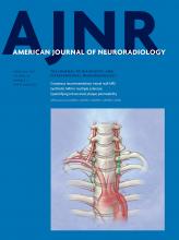
- Fig 1.
The “white gray sign.” Axial high-resolution 3D inversion recovery fast-spoiled gradient-echo T1-weighted image demonstrates decreased gray-white contrast of the anterior and posterior cortices along the central sulcus (white arrow).
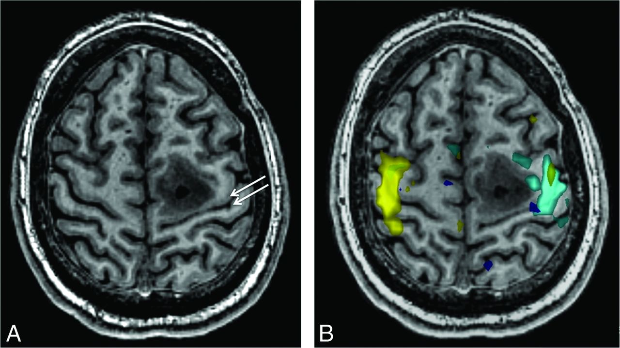
- Fig 2.
Measurement of gray-white contrast. A, Cortical segmentations were drawn along both sides of the central sulcus (royal blue and yellow) as well as along the adjacent banks of the precentral and postcentral sulci (red and pink) and the intervening WM (green and light blue). B, Gray-white contrast (± standard deviation bars) along the central sulcus was significantly (P < .001) decreased both anteriorly and posteriorly compared with the contrast along the pre- and postcentral sulci, respectively.
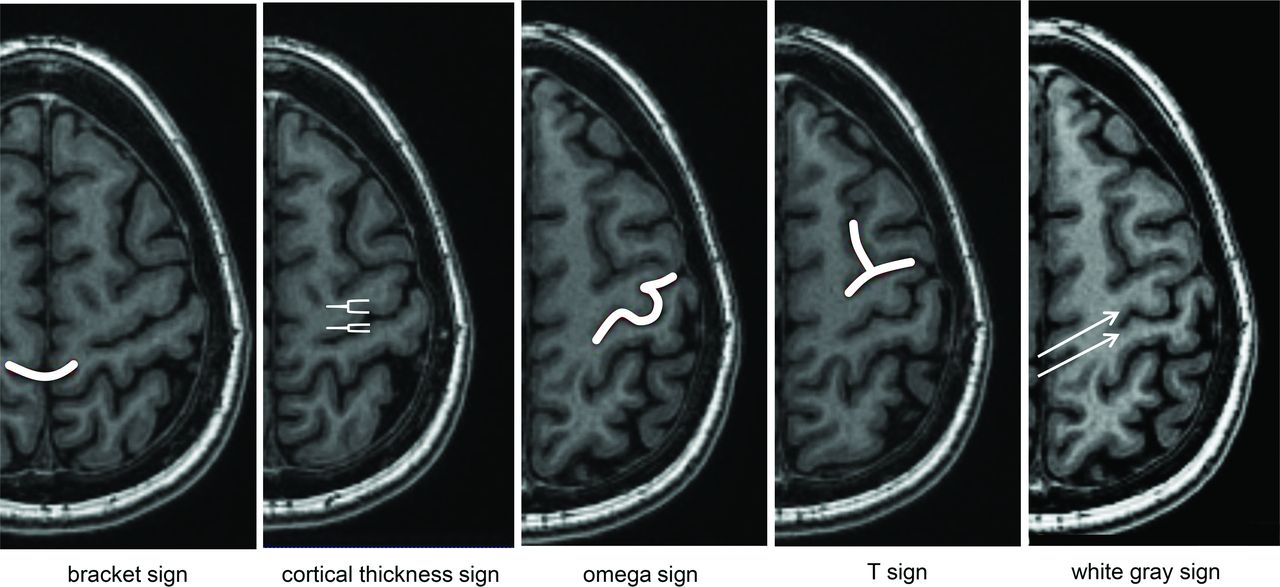
- Fig 3.
Signs of the central sulcus rated in this study. Bracket = central sulcus points to the marginal sulcus. Cortical thickness = increased cortical thickness along the anterior compared with posterior bank of the central sulcus. Omega = characteristic omega shape of the hand-motor knob. T = superior frontal sulcus meets the precentral gyrus.
- Fig 4.
Rater evaluation of signs of the central sulcus in 51 hemispheres (35 normal, 16 with pathology). 1 = definitely not present, 2 = likely not present, 3 = likely present, 4 = definitely present.
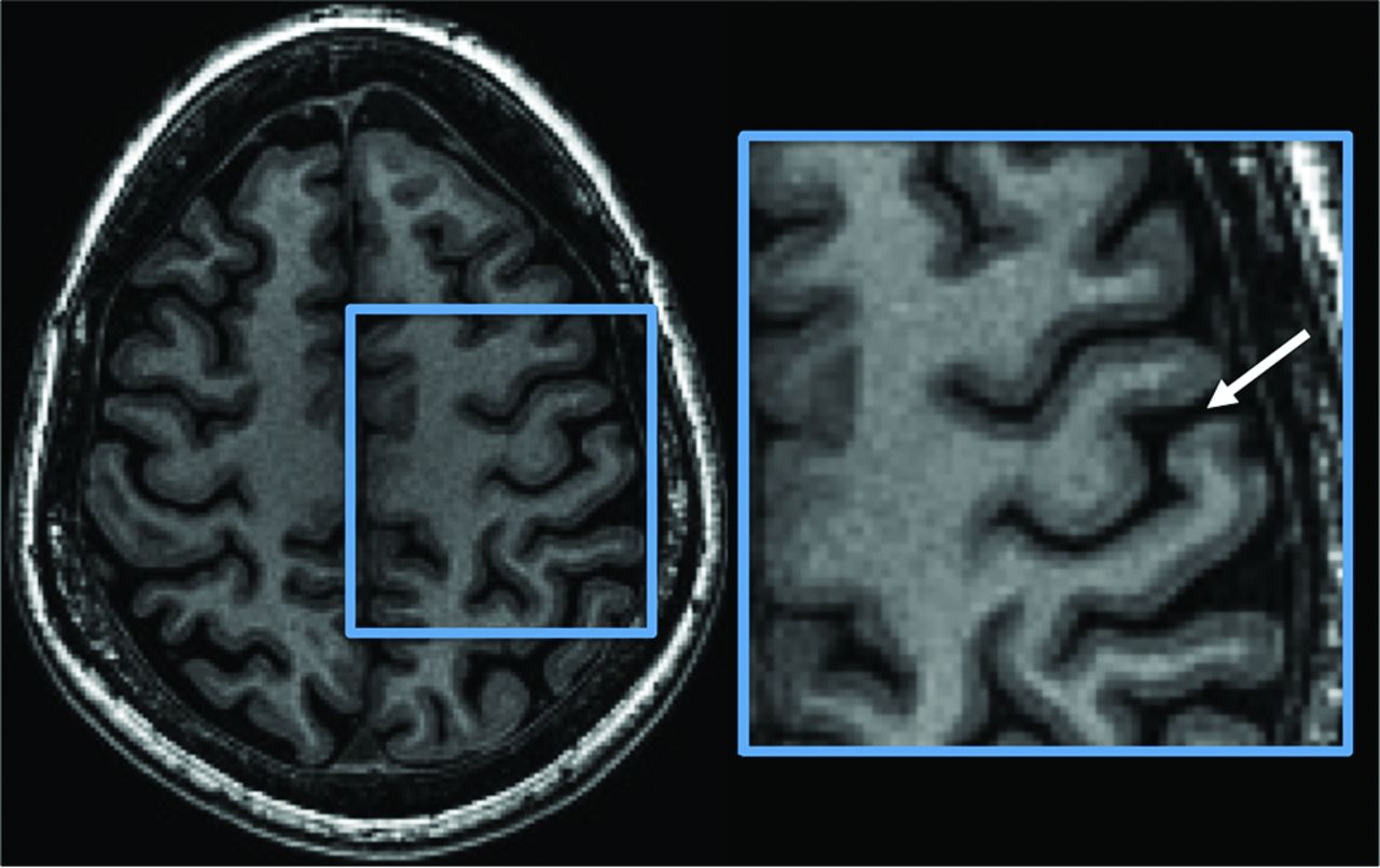
- Fig 5.
A 22-year-old man with posterior left frontal grade 2 astrocytoma. A, Axial 3D inversion recovery fast-spoiled gradient-echo T1-weighted image shows the tumor centered along the anterior aspect of the precentral gyrus. The white gray sign is still noticeable as decreased contrast of the gray-white interface along the central sulcus (arrows). This is also appreciable on the contralateral normal side. B, Functional data with hand-motor tasks (light blue = right hand, yellow = left hand) confirm the location of the primary motor and somatosensory cortices.


Tables
Comparison of the white gray sign with other signs of the central sulcusa
Hemispheres White Gray Bracket Thickness Omega T Rater 1 51 45 11 (<.001)b 23 (<.001)b 37 (.022) 38 (.092) 35 normal 34 9 (<.001)b 15 (<.001)b 26 28 16 abnormal 11 2 (.004)b 8 (.375) 11 10 Rater 2 51 51 38 (<.001)b 49 (.500) 42 (.004)b 27 (<.001)b 35 normal 35 28 (.016)b 35 28 (.016)b 20 (<.001)b 16 abnormal 16 10 (.031) 14 14 (.500) 7 (<.004)b ↵a Proportion of cases where each sign was reported, each compared with the white gray sign within rater. For the comparison of both hemispheres, a Bonferroni-adjusted P value of <.0125 was statistically significant correcting for the 4 signs evaluated. For the post-hoc separate evaluation of normal and abnormal hemispheres, the corrected threshold is P < .025.
↵b Statistically significant.


Jump to section
Related Articles
Cited By...
- No citing articles found.