
Article Figures & Data
Figures
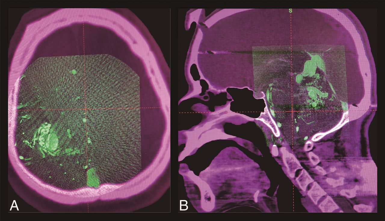
- Fig 1.
3D rotational angiography images obtained with a large (A) and an 8-inch detector (B) are coregistered to each stereotactic CT image on treatment-planning software (GammaPlan).
- Fig 2.
A 34-year-old woman with an unruptured, small, left medial frontal arteriovenous malformation, once treated with gamma knife (A). The small remnant nidus persisted at 3.5 years from the initial radiosurgery (B, a red arrowhead shows the remnant nidus). The actual treatment planning of the secondary treatment (C) shows that 3D rotational angiography (left column) successfully depicts the faint remnant, while both time-of-flight (middle column) and gadolinium-enhanced T1 images (right column) fail to depict it. The nidus was finally obliterated (D) at 1.5 years from the secondary treatment. Yellow lines show the prescription isodose lines.


Jump to section
Related Articles
Cited By...
- No citing articles found.