Advertisement

Main menu

  • Home
  • Content
    • Current Issue
    • Accepted Manuscripts
    • Article Preview
    • Past Issue Archive
    • Video Articles
    • AJNR Case Collection
    • Case of the Week Archive
    • Case of the Month Archive
    • Classic Case Archive
  • Special Collections
    • AJNR Awards
    • ASNR Foundation Special Collection
    • Most Impactful AJNR Articles
    • Photon-Counting CT
    • Spinal CSF Leak Articles (Jan 2020-June 2024)
  • Multimedia
    • AJNR Podcasts
    • AJNR SCANtastic
    • Trainee Corner
    • MRI Safety Corner
    • Imaging Protocols
  • For Authors
    • Submit a Manuscript
    • Submit a Video Article
    • Submit an eLetter to the Editor/Response
    • Manuscript Submission Guidelines
    • Statistical Tips
    • Fast Publishing of Accepted Manuscripts
    • Graphical Abstract Preparation
    • Imaging Protocol Submission
    • Author Policies
  • About Us
    • About AJNR
    • Editorial Board
    • Editorial Board Alumni
  • More
    • Become a Reviewer/Academy of Reviewers
    • Subscribers
    • Permissions
    • Alerts
    • Feedback
    • Advertisers
    • ASNR Home

User menu

  • Alerts
  • Log in

Search

  • Advanced search
American Journal of Neuroradiology

American Journal of Neuroradiology

ASHNR American Society of Functional Neuroradiology ASHNR American Society of Pediatric Neuroradiology ASSR
  • Alerts
  • Log in

Advanced Search

  • Home
  • Content
    • Current Issue
    • Accepted Manuscripts
    • Article Preview
    • Past Issue Archive
    • Video Articles
    • AJNR Case Collection
    • Case of the Week Archive
    • Case of the Month Archive
    • Classic Case Archive
  • Special Collections
    • AJNR Awards
    • ASNR Foundation Special Collection
    • Most Impactful AJNR Articles
    • Photon-Counting CT
    • Spinal CSF Leak Articles (Jan 2020-June 2024)
  • Multimedia
    • AJNR Podcasts
    • AJNR SCANtastic
    • Trainee Corner
    • MRI Safety Corner
    • Imaging Protocols
  • For Authors
    • Submit a Manuscript
    • Submit a Video Article
    • Submit an eLetter to the Editor/Response
    • Manuscript Submission Guidelines
    • Statistical Tips
    • Fast Publishing of Accepted Manuscripts
    • Graphical Abstract Preparation
    • Imaging Protocol Submission
    • Author Policies
  • About Us
    • About AJNR
    • Editorial Board
    • Editorial Board Alumni
  • More
    • Become a Reviewer/Academy of Reviewers
    • Subscribers
    • Permissions
    • Alerts
    • Feedback
    • Advertisers
    • ASNR Home
  • Follow AJNR on Twitter
  • Visit AJNR on Facebook
  • Follow AJNR on Instagram
  • Join AJNR on LinkedIn
  • RSS Feeds

AJNR is seeking candidates for the AJNR Podcast Editor. Read the position description.

Research ArticleHead & Neck

Facial Venous Malformations Are Associated with Cerebral Developmental Venous Anomalies

W. Brinjikji, C.A. Hilditch, A.C. Tsang, P.J. Nicholson, T. Krings and R. Agid
American Journal of Neuroradiology November 2018, 39 (11) 2103-2107; DOI: https://doi.org/10.3174/ajnr.A5811
W. Brinjikji
aFrom the Departments of Radiology (W.B.)
bNeurosurgery (W.B.), Mayo Clinic, Rochester, Minnesota
cJoint Department of Medical Imaging (W.B., C.A.H., A.C.T., P.J.N., T.K., R.A.), Toronto Western Hospital, Toronto, Ontario, Canada.
  • Find this author on Google Scholar
  • Find this author on PubMed
  • Search for this author on this site
  • ORCID record for W. Brinjikji
C.A. Hilditch
cJoint Department of Medical Imaging (W.B., C.A.H., A.C.T., P.J.N., T.K., R.A.), Toronto Western Hospital, Toronto, Ontario, Canada.
  • Find this author on Google Scholar
  • Find this author on PubMed
  • Search for this author on this site
  • ORCID record for C.A. Hilditch
A.C. Tsang
cJoint Department of Medical Imaging (W.B., C.A.H., A.C.T., P.J.N., T.K., R.A.), Toronto Western Hospital, Toronto, Ontario, Canada.
  • Find this author on Google Scholar
  • Find this author on PubMed
  • Search for this author on this site
  • ORCID record for A.C. Tsang
P.J. Nicholson
cJoint Department of Medical Imaging (W.B., C.A.H., A.C.T., P.J.N., T.K., R.A.), Toronto Western Hospital, Toronto, Ontario, Canada.
  • Find this author on Google Scholar
  • Find this author on PubMed
  • Search for this author on this site
  • ORCID record for P.J. Nicholson
T. Krings
cJoint Department of Medical Imaging (W.B., C.A.H., A.C.T., P.J.N., T.K., R.A.), Toronto Western Hospital, Toronto, Ontario, Canada.
  • Find this author on Google Scholar
  • Find this author on PubMed
  • Search for this author on this site
  • ORCID record for T. Krings
R. Agid
cJoint Department of Medical Imaging (W.B., C.A.H., A.C.T., P.J.N., T.K., R.A.), Toronto Western Hospital, Toronto, Ontario, Canada.
  • Find this author on Google Scholar
  • Find this author on PubMed
  • Search for this author on this site
  • ORCID record for R. Agid
  • Article
  • Figures & Data
  • Supplemental
  • Info & Metrics
  • Responses
  • References
  • PDF
Loading

Article Figures & Data

Figures

  • Fig 1.
    • Download figure
    • Open in new tab
    • Download powerpoint
    Fig 1.

    A 49-year-old woman with VMs and DVAs. VMs are in the left temporal region, orbit, zygomaticofacial region, and mandible (A and B). The patient underwent bleomycin sclerotherapy for treatment of the VMs with good results (C). She also had an extensive DVA of the left temporal lobe, basal ganglia, and left cerebellar hemisphere (D and E). Findings would be consistent with CVMS 1–3.
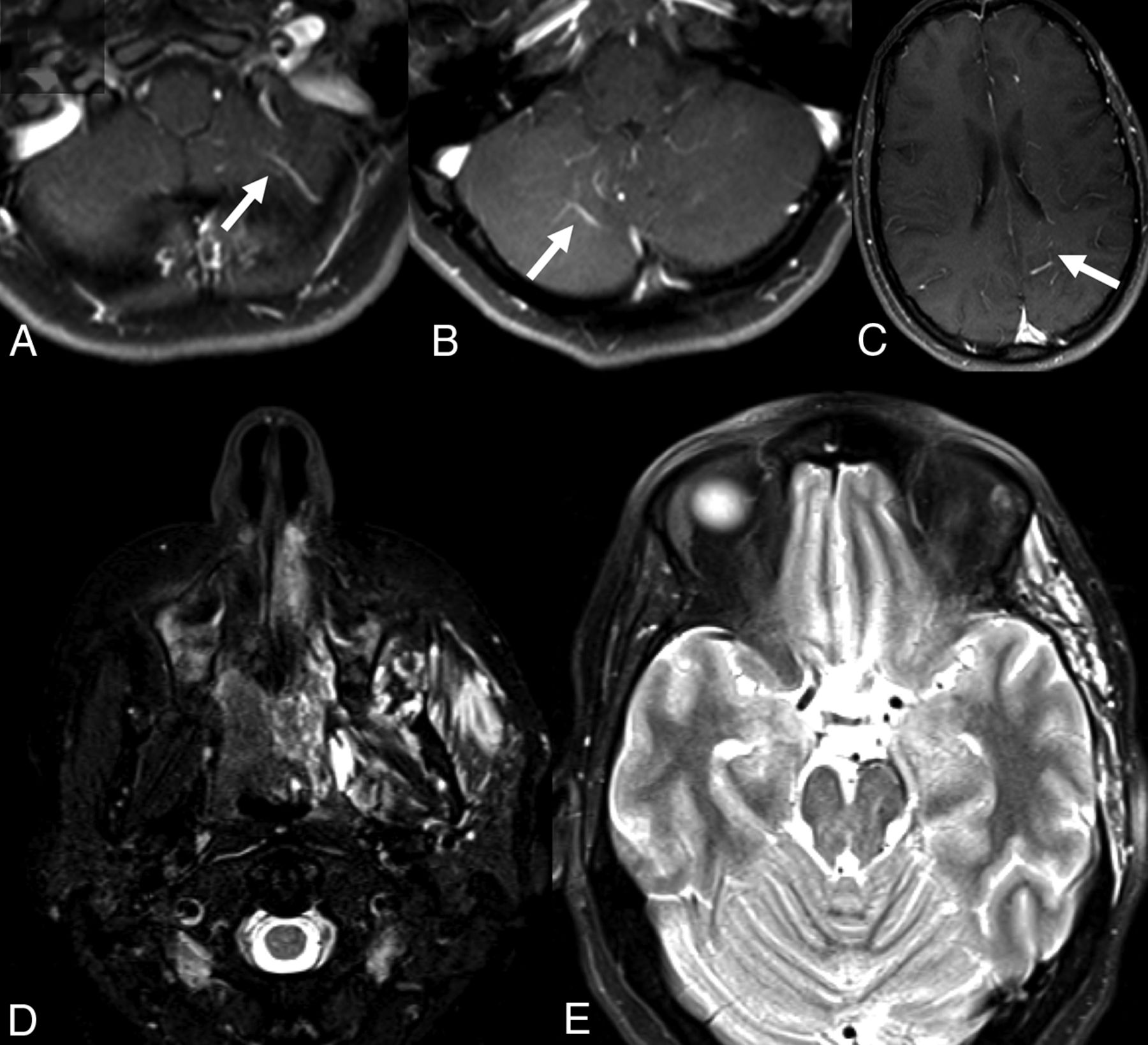
  • Fig 2.
    • Download figure
    • Open in new tab
    • Download powerpoint
    Fig 2.

    A 30-year-old man with facial VMs and left supratentorial DVAs. A–C, Postcontrast T1-weighted MRIs show DVAs in the bilateral cerebellar hemispheres, left parietal lobe, and left temporal lobe. D and E, T2-weighted MR imaging shows extensive VMs of the left zygomaticotemporal region and masticator spaces.
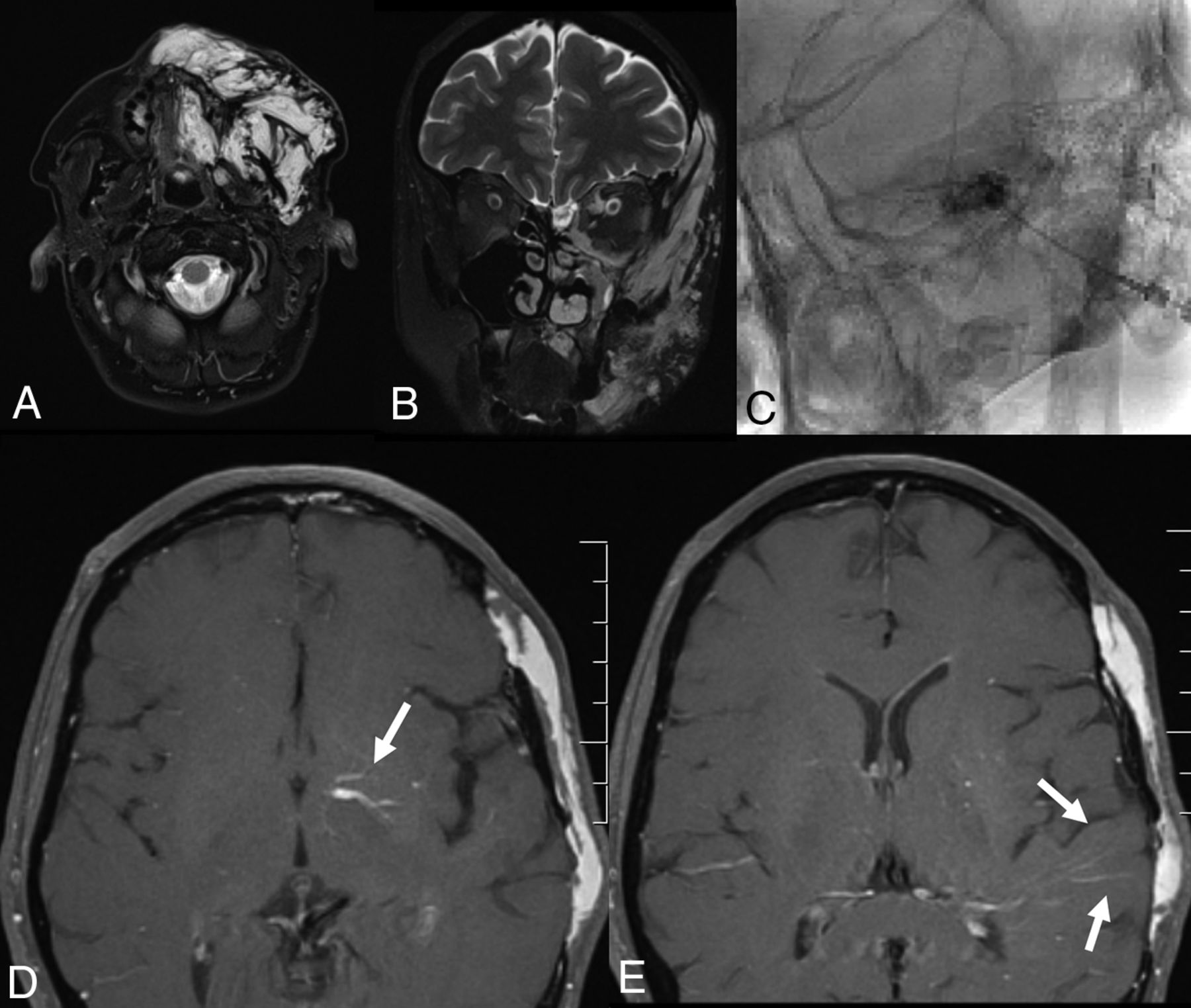
  • Fig 3.
    • Download figure
    • Open in new tab
    • Download powerpoint
    Fig 3.

    A 22-year-old woman with a left facial DVA, bilateral VMs, and a de novo cavernoma. A, Coronal T2-weighted MR imaging demonstrates a vascular malformation involving the soft tissues overlying the left zygomatico-orbital region with extension to the left maxillary region. B, Axial SWI MR imaging of the brain demonstrates extensive DVAs of the bilateral cerebellar hemispheres and a cavernoma of the left medulla. C, Postcontrast MR imaging demonstrates venous radicles of 2 DVAs involving the bilateral basal ganglia. D, T2-weighted MR imaging 2 years later demonstrates a large cavernoma that developed in one of the venous radicles of the left basal ganglia DVA.

PreviousNext
Back to top

In this issue

American Journal of Neuroradiology: 39 (11)
American Journal of Neuroradiology
Vol. 39, Issue 11
1 Nov 2018
  • Table of Contents
  • Index by author
  • Complete Issue (PDF)
Advertisement
Print
Download PDF
Email Article

Thank you for your interest in spreading the word on American Journal of Neuroradiology.

NOTE: We only request your email address so that the person you are recommending the page to knows that you wanted them to see it, and that it is not junk mail. We do not capture any email address.

Enter multiple addresses on separate lines or separate them with commas.
Facial Venous Malformations Are Associated with Cerebral Developmental Venous Anomalies
(Your Name) has sent you a message from American Journal of Neuroradiology
(Your Name) thought you would like to see the American Journal of Neuroradiology web site.
CAPTCHA
This question is for testing whether or not you are a human visitor and to prevent automated spam submissions.
Cite this article
W. Brinjikji, C.A. Hilditch, A.C. Tsang, P.J. Nicholson, T. Krings, R. Agid
Facial Venous Malformations Are Associated with Cerebral Developmental Venous Anomalies
American Journal of Neuroradiology Nov 2018, 39 (11) 2103-2107; DOI: 10.3174/ajnr.A5811

Citation Manager Formats

  • BibTeX
  • Bookends
  • EasyBib
  • EndNote (tagged)
  • EndNote 8 (xml)
  • Medlars
  • Mendeley
  • Papers
  • RefWorks Tagged
  • Ref Manager
  • RIS
  • Zotero
0 Responses
Respond to this article
Share
Bookmark this article
Facial Venous Malformations Are Associated with Cerebral Developmental Venous Anomalies
W. Brinjikji, C.A. Hilditch, A.C. Tsang, P.J. Nicholson, T. Krings, R. Agid
American Journal of Neuroradiology Nov 2018, 39 (11) 2103-2107; DOI: 10.3174/ajnr.A5811
del.icio.us logo Twitter logo Facebook logo Mendeley logo
  • Tweet Widget
  • Facebook Like
  • Google Plus One
Purchase

Jump to section

  • Article
    • Abstract
    • ABBREVIATIONS:
    • Materials and Methods
    • Results
    • Discussion
    • Conclusions
    • References
  • Figures & Data
  • Supplemental
  • Info & Metrics
  • Responses
  • References
  • PDF

Related Articles

  • PubMed
  • Google Scholar

Cited By...

  • Cerebrofacial vascular metameric syndrome is caused by somatic pathogenic variants in PIK3CA
  • Developmental Venous Anomalies are a Genetic Primer for Cerebral Cavernous Malformations
  • Cervicofacial Venous Malformations Are Associated with Intracranial Developmental Venous Anomalies and Dural Venous Sinus Abnormalities
  • Crossref (18)
  • Google Scholar

This article has been cited by the following articles in journals that are participating in Crossref Cited-by Linking.

  • Developmental venous anomalies are a genetic primer for cerebral cavernous malformations
    Daniel A. Snellings, Romuald Girard, Rhonda Lightle, Abhinav Srinath, Sharbel Romanos, Ying Li, Chang Chen, Aileen A. Ren, Mark L. Kahn, Issam A. Awad, Douglas A. Marchuk
    Nature Cardiovascular Research 2022 1 3
  • Cerebrofacial venous metameric syndrome—spectrum of imaging findings
    Waleed Brinjikji, Patrick Nicholson, Christopher A Hilditch, Timo Krings, Vitor Pereira, Ronit Agid
    Neuroradiology 2020 62 4
  • Venous pathologies in paediatric neuroradiology: from foetal to adolescent life
    Kshitij Mankad, Asthik Biswas, Maria Camilla Rossi Espagnet, Luke Dixon, Nihaal Reddy, Ai Peng Tan, Ozgur Oztekin, Felice D’Arco, Karuna Shekdar, Prakash Muthusami, Fergus Robertson, Stacy Goergen, Winston Chong
    Neuroradiology 2020 62 1
  • Symptomatic developmental venous anomalies
    Lorenzo Rinaldo, Giuseppe Lanzino, Kelly D. Flemming, Timo Krings, Waleed Brinjikji
    Acta Neurochirurgica 2020 162 5
  • High Prevalence of Developmental Venous Anomaly in Diffuse Intrinsic Pontine Gliomas: A Pediatric Control Study
    Alexandre Roux, Nathalie Boddaert, Jacques Grill, David Castel, Marc Zanello, Gilles Zah-Bi, Fabrice Chrétien, Etienne Lefevre, Volodia Dangouloff Ros, Michel Zerah, Stéphanie Puget, Johan Pallud, Pascale Varlet
    Neurosurgery 2020 86 4
  • Cervicofacial Venous Malformations Are Associated with Intracranial Developmental Venous Anomalies and Dural Venous Sinus Abnormalities
    W. Brinjikji, I.T. Mark, V.M. Silvera, J.B. Guerin
    American Journal of Neuroradiology 2020 41 7
  • Intracranial venous malformations: Incidence and characterization in a large pediatric cohort
    Karen S Chen, Alaa Montaser, Ramsey Ashour, Darren B Orbach
    Interventional Neuroradiology 2021 27 1
  • Cerebrofacial vascular metameric syndrome is caused by somatic pathogenic variants in PIK3CA
    Sarah E. Sheppard, Victoria R. Sanders, Abhay Srinivasan, Laura S. Finn, Denise Adams, Andrew Elton, Catherine Amlie-Lefond, Zoe Nelson, Victoria Dmyterko, Dana Jensen, Kaitlyn Zenner, Jonathan Perkins, James T. Bennett
    Molecular Case Studies 2021 7 6
  • Intracranial and extracranial vascular manifestations of patients with a clinical diagnosis of Klippel–Trenaunay syndrome
    Tristan N. Covington, Katelyn R. Anderson, Megha M. Tollefson, Julie B. Guerin, Waleed Brinjikji
    Neuroradiology 2021 63 3
  • Letter: Is Developmental Venous Anomaly an Imaging Biomarker of PIK3CA Mutated Gliomas?
    Alexandre Roux, Miikka Vikkula, Johan Pallud
    Neurosurgery 2020 86 1

More in this TOC Section

  • WHO Classification Update: Nasal&Skull Base Tumors
  • Peritumoral Signal in Vestibular Schwannomas
  • NI-RADS for HEAD&NECK Cancer Recurrence
Show more HEAD & NECK

Similar Articles

Advertisement

Indexed Content

  • Current Issue
  • Accepted Manuscripts
  • Article Preview
  • Past Issues
  • Editorials
  • Editor's Choice
  • Fellows' Journal Club
  • Letters to the Editor
  • Video Articles

Cases

  • Case Collection
  • Archive - Case of the Week
  • Archive - Case of the Month
  • Archive - Classic Case

Special Collections

  • AJNR Awards
  • ASNR Foundation Special Collection
  • Most Impactful AJNR Articles
  • Photon-Counting CT
  • Spinal CSF Leak Articles (Jan 2020-June 2024)

More from AJNR

  • Trainee Corner
  • Imaging Protocols
  • MRI Safety Corner

Multimedia

  • AJNR Podcasts
  • AJNR Scantastics

Resources

  • Turnaround Time
  • Submit a Manuscript
  • Submit a Video Article
  • Submit an eLetter to the Editor/Response
  • Manuscript Submission Guidelines
  • Statistical Tips
  • Fast Publishing of Accepted Manuscripts
  • Graphical Abstract Preparation
  • Imaging Protocol Submission
  • Evidence-Based Medicine Level Guide
  • Publishing Checklists
  • Author Policies
  • Become a Reviewer/Academy of Reviewers
  • News and Updates

About Us

  • About AJNR
  • Editorial Board
  • Editorial Board Alumni
  • Alerts
  • Permissions
  • Not an AJNR Subscriber? Join Now
  • Advertise with Us
  • Librarian Resources
  • Feedback
  • Terms and Conditions
  • AJNR Editorial Board Alumni

American Society of Neuroradiology

  • Not an ASNR Member? Join Now

© 2025 by the American Society of Neuroradiology All rights, including for text and data mining, AI training, and similar technologies, are reserved.
Print ISSN: 0195-6108 Online ISSN: 1936-959X

Powered by HighWire