
Article Figures & Data
Figures
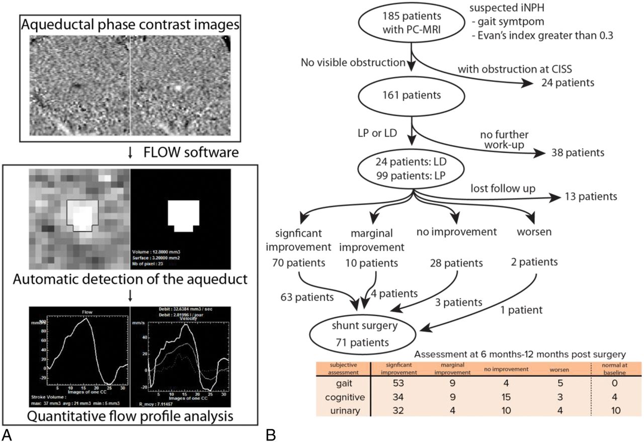
- Fig 1.
An overview of the AQ-PCMR image-processing pipeline and treatment flowchart of patients suspected of having CAH in this study. A, The phase-contrast MR imaging data acquired at the level of cerebral aqueduct are analyzed by Flow software to calculate the stroke volume and flow rate of each patient. B, One hundred eighty-five patients with gait abnormality and ventricular enlargement determined by the Evans index are included in this study. Depending on quantitative and subjective response to LP/LD, patients undergo shunt surgery. The shunt outcomes are determined within 12 months after surgery using both subjective assessment in 3 symptom categories and quantitative measurements such as the Tinetti, TUG, or MMSE scores.
- Fig 2.
Quantitative assessment for LP/LD and shunt response. A, Comparison between the improvement in TUG scores after LP/LD (x-axis) and after shunt surgery (y-axis). Thirty-one patients show better improvement after shunt surgery than after LP/LD, whereas 5 patients show better improvement after LP/LD than after shunt surgery. B, Comparison between the improvement in the Tinetti score “deficit” after LP/LD (x-axis) and after shunt surgery (y-axis). Patients with a Tinetti score of 28 at baseline and 2 outliers (−1.4, 0.6) and (−1, −2) are not shown on this graph. Thirty-one patients show better improvement after shunt surgery than after LP/LD, whereas 9 patients show better improvement after LP/LD than after shunt surgery. C, Changes in the MMSE score after shunt surgery. Blue lines represent patients who improved, and red lines represent patients who worsened by ≥3 points in the MMSE test after shunt surgery.
- Fig 3.
The aqueductal CSF flow and LP/LD response measured by subjective improvement and Tinetti and TUG score improvement. A, The distributions of aqueductal stroke volume (upper) and flow rate (lower) are compared between LP/LD responders and nonresponders. The aqueductal flow rate is significantly higher in LP/LD nonresponders than in responders (Wilcoxon rank sum test, P = .03 for stroke volume and .028 for flow rate). B, The aqueductal stroke volume (upper) and flow rate (lower) are plotted against the quantitative improvement of the TUG score (y-axis) after LP/LD. The Pearson correlation between the TUG score improvement and the stroke volume and flow rate are −0.113 and −0.116, respectively. C, Twenty percent or greater improvement in the TUG score after LP/LD was used to define LP/LD responders, and <20% improvement in the TUG score after LP/LD was used to define LP/LD nonresponders. The number of patients in each group is shown as a number on the graph. The 2 groups exhibit an overlapping distribution of aqueductal flow rate and stroke volumes. D, “Tinetti score deficit” is defined as the difference between a perfect Tinetti score and the patient's Tinetti score. Twenty percent or greater improvement in the Tinetti score deficit after LP/LD was used to define LP/LD responders, and < 20% improvement in the Tinetti score deficit after LP/LD was used to define LP/LD nonresponders. Again, the 2 groups exhibit overlapping distribution of aqueductal flow rate and stroke volumes.


