Advertisement

Main menu

  • Home
  • Content
    • Current Issue
    • Accepted Manuscripts
    • Article Preview
    • Past Issue Archive
    • Video Articles
    • AJNR Case Collection
    • Case of the Week Archive
    • Case of the Month Archive
    • Classic Case Archive
  • Special Collections
    • AJNR Awards
    • ASNR Foundation Special Collection
    • Most Impactful AJNR Articles
    • Photon-Counting CT
    • Spinal CSF Leak Articles (Jan 2020-June 2024)
  • Multimedia
    • AJNR Podcasts
    • AJNR SCANtastic
    • Trainee Corner
    • MRI Safety Corner
    • Imaging Protocols
  • For Authors
    • Submit a Manuscript
    • Submit a Video Article
    • Submit an eLetter to the Editor/Response
    • Manuscript Submission Guidelines
    • Statistical Tips
    • Fast Publishing of Accepted Manuscripts
    • Graphical Abstract Preparation
    • Imaging Protocol Submission
    • Author Policies
  • About Us
    • About AJNR
    • Editorial Board
    • Editorial Board Alumni
  • More
    • Become a Reviewer/Academy of Reviewers
    • Subscribers
    • Permissions
    • Alerts
    • Feedback
    • Advertisers
    • ASNR Home

User menu

  • Alerts
  • Log in

Search

  • Advanced search
American Journal of Neuroradiology

American Journal of Neuroradiology

ASHNR American Society of Functional Neuroradiology ASHNR American Society of Pediatric Neuroradiology ASSR
  • Alerts
  • Log in

Advanced Search

  • Home
  • Content
    • Current Issue
    • Accepted Manuscripts
    • Article Preview
    • Past Issue Archive
    • Video Articles
    • AJNR Case Collection
    • Case of the Week Archive
    • Case of the Month Archive
    • Classic Case Archive
  • Special Collections
    • AJNR Awards
    • ASNR Foundation Special Collection
    • Most Impactful AJNR Articles
    • Photon-Counting CT
    • Spinal CSF Leak Articles (Jan 2020-June 2024)
  • Multimedia
    • AJNR Podcasts
    • AJNR SCANtastic
    • Trainee Corner
    • MRI Safety Corner
    • Imaging Protocols
  • For Authors
    • Submit a Manuscript
    • Submit a Video Article
    • Submit an eLetter to the Editor/Response
    • Manuscript Submission Guidelines
    • Statistical Tips
    • Fast Publishing of Accepted Manuscripts
    • Graphical Abstract Preparation
    • Imaging Protocol Submission
    • Author Policies
  • About Us
    • About AJNR
    • Editorial Board
    • Editorial Board Alumni
  • More
    • Become a Reviewer/Academy of Reviewers
    • Subscribers
    • Permissions
    • Alerts
    • Feedback
    • Advertisers
    • ASNR Home
  • Follow AJNR on Twitter
  • Visit AJNR on Facebook
  • Follow AJNR on Instagram
  • Join AJNR on LinkedIn
  • RSS Feeds

AJNR is seeking candidates for the AJNR Podcast Editor. Read the position description.

Research ArticlePediatrics

Using Correlative Properties of Neighboring Pixels to Improve Gray-White Differentiation in Pediatric Head CT Images

T.P. Madaelil, A. Sharma, C. Hildebolt and M. Parsons
American Journal of Neuroradiology March 2018, 39 (3) 577-582; DOI: https://doi.org/10.3174/ajnr.A5506
T.P. Madaelil
aFrom the Department of Interventional Neuroradiology (T.P.M.), Emory University Hospital, Atlanta, Georgia
  • Find this author on Google Scholar
  • Find this author on PubMed
  • Search for this author on this site
  • ORCID record for T.P. Madaelil
A. Sharma
bMallinckrodt Institute of Radiology (A.S., C.H., M.P.), Washington University School of Medicine, Washington University, St. Louis, Missouri.
  • Find this author on Google Scholar
  • Find this author on PubMed
  • Search for this author on this site
  • ORCID record for A. Sharma
C. Hildebolt
bMallinckrodt Institute of Radiology (A.S., C.H., M.P.), Washington University School of Medicine, Washington University, St. Louis, Missouri.
  • Find this author on Google Scholar
  • Find this author on PubMed
  • Search for this author on this site
  • ORCID record for C. Hildebolt
M. Parsons
bMallinckrodt Institute of Radiology (A.S., C.H., M.P.), Washington University School of Medicine, Washington University, St. Louis, Missouri.
  • Find this author on Google Scholar
  • Find this author on PubMed
  • Search for this author on this site
  • ORCID record for M. Parsons
  • Article
  • Figures & Data
  • Info & Metrics
  • Responses
  • References
  • PDF
Loading

Article Figures & Data

Figures

  • Fig 1.
    • Download figure
    • Open in new tab
    • Download powerpoint
    Fig 1.

    These images represent 9 contiguous pixels representing the same structure that spans these pixels. In an ideal noise-free image (A), these 9 pixels would have identical intensities. The presence of noise that invariably accompanies digital images causes intensities of some of these pixels to be higher or lower than expected (B). Modifying the intensities of pixels (asterisk) on the basis of the preponderance of intensities (plus sign) in their neighboring pixels can mitigate the effect of noise (C).

  • Fig 2.
    • Download figure
    • Open in new tab
    • Download powerpoint
    Fig 2.

    Axial head CT image across the centrum semiovale before (baseline image) and after (enhanced image) processing with CIE, with equivalent ROIs used to measure gray and white matter intensities. These measurements were used to calculate the conspicuity of gray matter and CNR between gray and white matter.
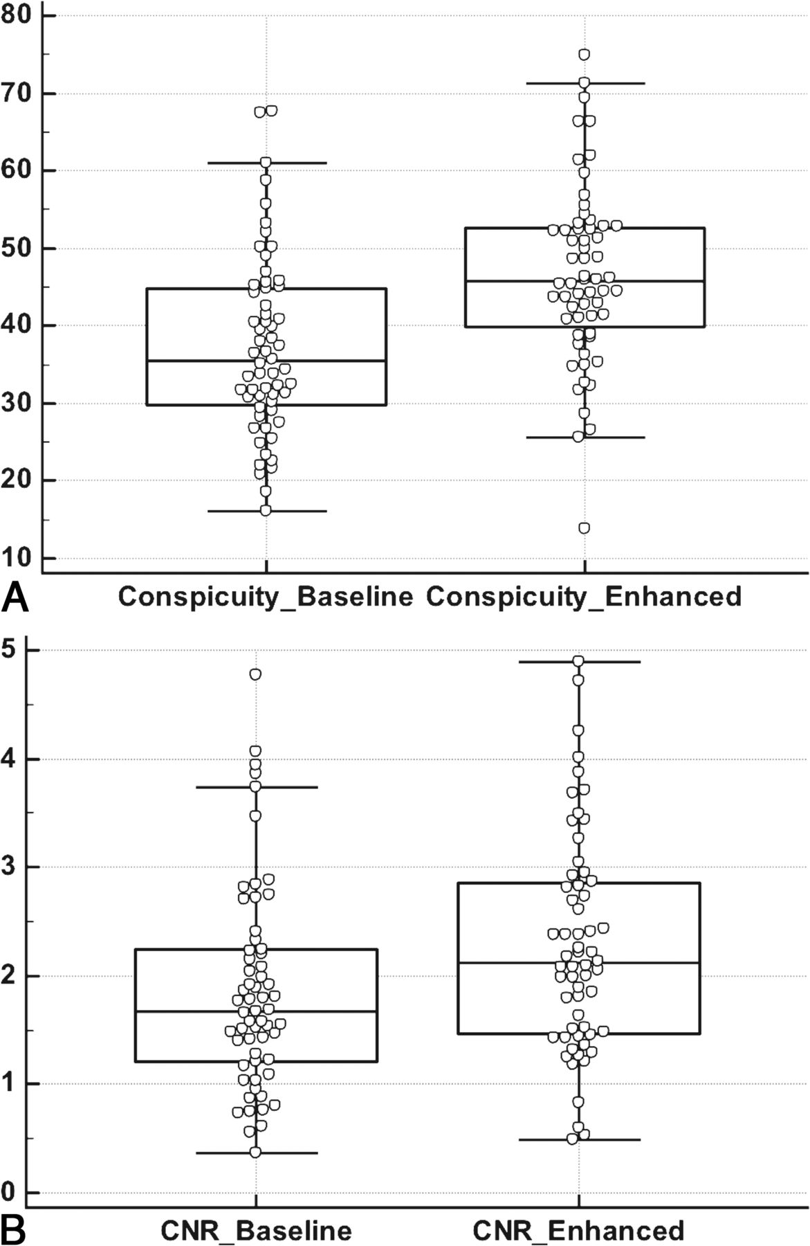
  • Fig 3.
    • Download figure
    • Open in new tab
    • Download powerpoint
    Fig 3.

    Boxplots showing the distribution of gray matter conspicuity (A) and CNR between gray and white matter (B) in 60 paired head CT images with normal findings before (baseline) and after (enhanced) processing with CIE. Both of these quantitative measures of gray-white differentiation demonstrated significant improvement following image processing (P < .001 for conspicuity; P = .015 for CNR).

  • Fig 4.
    • Download figure
    • Open in new tab
    • Download powerpoint
    Fig 4.

    Clustered columns showing the distribution of qualitative GWD ratings assigned by 2 blinded radiologists to baseline and enhanced images on a 5-point Likert scale ranging from 1 (imperceptible GWD) to 5 (very easily perceptible GWD). The vertical axis shows the percentage of all cases assigned a given GWD rating. A favorable shift toward higher GWD ratings was seen with image processing for both radiologists (P < .01).
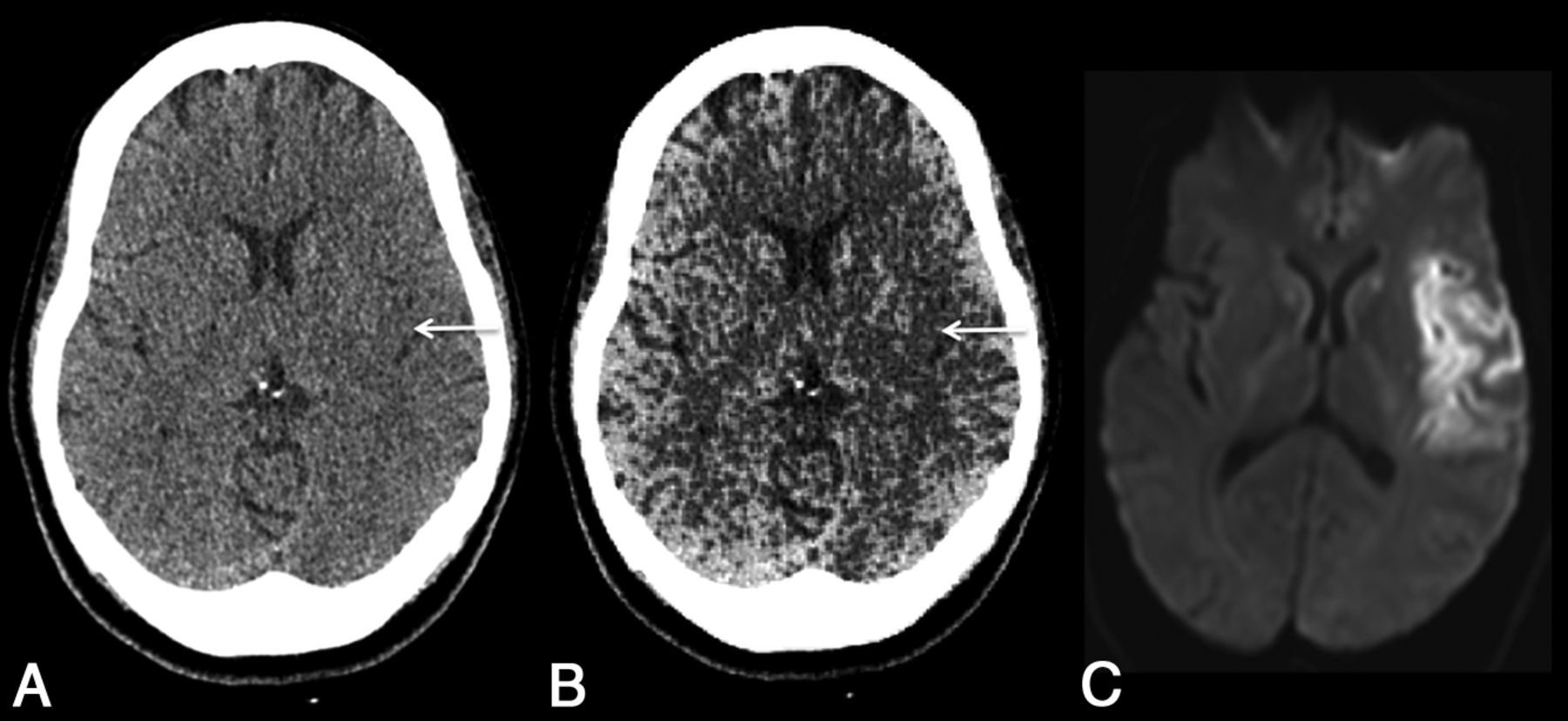
  • Fig 5.
    • Download figure
    • Open in new tab
    • Download powerpoint
    Fig 5.

    Axial head CT image across the insula obtained within 3 hours of stroke onset before (A) and after (B) processing with CIE. Note that improved gray-white differentiation following image processing makes it easier to perceive the loss of normal gray matter density in the insula (arrow), corresponding to the infarction proved on subsequent DWI (C).

PreviousNext
Back to top

In this issue

American Journal of Neuroradiology: 39 (3)
American Journal of Neuroradiology
Vol. 39, Issue 3
1 Mar 2018
  • Table of Contents
  • Index by author
  • Complete Issue (PDF)
Advertisement
Print
Download PDF
Email Article

Thank you for your interest in spreading the word on American Journal of Neuroradiology.

NOTE: We only request your email address so that the person you are recommending the page to knows that you wanted them to see it, and that it is not junk mail. We do not capture any email address.

Enter multiple addresses on separate lines or separate them with commas.
Using Correlative Properties of Neighboring Pixels to Improve Gray-White Differentiation in Pediatric Head CT Images
(Your Name) has sent you a message from American Journal of Neuroradiology
(Your Name) thought you would like to see the American Journal of Neuroradiology web site.
CAPTCHA
This question is for testing whether or not you are a human visitor and to prevent automated spam submissions.
Cite this article
T.P. Madaelil, A. Sharma, C. Hildebolt, M. Parsons
Using Correlative Properties of Neighboring Pixels to Improve Gray-White Differentiation in Pediatric Head CT Images
American Journal of Neuroradiology Mar 2018, 39 (3) 577-582; DOI: 10.3174/ajnr.A5506

Citation Manager Formats

  • BibTeX
  • Bookends
  • EasyBib
  • EndNote (tagged)
  • EndNote 8 (xml)
  • Medlars
  • Mendeley
  • Papers
  • RefWorks Tagged
  • Ref Manager
  • RIS
  • Zotero
0 Responses
Respond to this article
Share
Bookmark this article
Using Correlative Properties of Neighboring Pixels to Improve Gray-White Differentiation in Pediatric Head CT Images
T.P. Madaelil, A. Sharma, C. Hildebolt, M. Parsons
American Journal of Neuroradiology Mar 2018, 39 (3) 577-582; DOI: 10.3174/ajnr.A5506
del.icio.us logo Twitter logo Facebook logo Mendeley logo
  • Tweet Widget
  • Facebook Like
  • Google Plus One
Purchase

Jump to section

  • Article
    • Abstract
    • ABBREVIATIONS:
    • Materials and Methods
    • Results
    • Discussion
    • Conclusions
    • Footnotes
    • References
  • Figures & Data
  • Info & Metrics
  • Responses
  • References
  • PDF

Related Articles

  • No related articles found.
  • PubMed
  • Google Scholar

Cited By...

  • Evaluating the Utility of a Postprocessing Algorithm for MRI Evaluation of Optic Neuritis
  • Crossref (5)
  • Google Scholar

This article has been cited by the following articles in journals that are participating in Crossref Cited-by Linking.

  • Evaluating the Utility of a Postprocessing Algorithm for MRI Evaluation of Optic Neuritis
    L. Stunkel, A. Sharma, M.S. Parsons, A. Salter, G.P. Van Stavern
    American Journal of Neuroradiology 2019 40 6
  • Using Correlative Properties of Neighboring Pixels to Enhance Contrast-to-Noise Ratio of Abnormal Hippocampus in Patients With Intractable Epilepsy and Mesial Temporal Sclerosis
    Matthew S. Parsons, Aseem Sharma, Charles Hildebolt
    Academic Radiology 2019 26 4
  • Enhancing contrast to noise ratio of hippocampi affected with mesial temporal sclerosis: A case-control study in children undergoing epilepsy surgeries
    Hilary L.P. Orlowski, Matthew D. Smyth, Matthew S. Parsons, Sonika Dahiya, Nima Sharifai, Charles Hildebolt, Aseem Sharma
    Clinical Neurology and Neurosurgery 2018 174
  • Evaluating the effect of a post-processing algorithm in detection of annular fissure on MR imaging
    Rami W. Eldaya, Matthew S. Parsons, Hilary L. P. Orlowski, Martin N. Reis, Aseem Sharma
    European Spine Journal 2021 30 8
  • Delineation of Inner Annulus Fibrosus and Nucleus Pulposus on Routine T2-weighted MR Images
    Rami W. Eldaya, Matthew S. Parsons, Lee Rhea, Aseem Sharma
    Spine 2022 47 13

More in this TOC Section

  • SyMRI & MR Fingerprinting in Brainstem Myelination
  • Comparison of Image Quality and Radiation Dose in Pediatric Temporal Bone CT Using Photon-Counting Detector CT and Energy-Integrating Detector CT
  • Dual-Layer Detector CT for PEDS Image Quality
Show more Pediatrics

Similar Articles

Advertisement

Indexed Content

  • Current Issue
  • Accepted Manuscripts
  • Article Preview
  • Past Issues
  • Editorials
  • Editor's Choice
  • Fellows' Journal Club
  • Letters to the Editor
  • Video Articles

Cases

  • Case Collection
  • Archive - Case of the Week
  • Archive - Case of the Month
  • Archive - Classic Case

Special Collections

  • AJNR Awards
  • ASNR Foundation Special Collection
  • Most Impactful AJNR Articles
  • Photon-Counting CT
  • Spinal CSF Leak Articles (Jan 2020-June 2024)

More from AJNR

  • Trainee Corner
  • Imaging Protocols
  • MRI Safety Corner

Multimedia

  • AJNR Podcasts
  • AJNR Scantastics

Resources

  • Turnaround Time
  • Submit a Manuscript
  • Submit a Video Article
  • Submit an eLetter to the Editor/Response
  • Manuscript Submission Guidelines
  • Statistical Tips
  • Fast Publishing of Accepted Manuscripts
  • Graphical Abstract Preparation
  • Imaging Protocol Submission
  • Evidence-Based Medicine Level Guide
  • Publishing Checklists
  • Author Policies
  • Become a Reviewer/Academy of Reviewers
  • News and Updates

About Us

  • About AJNR
  • Editorial Board
  • Editorial Board Alumni
  • Alerts
  • Permissions
  • Not an AJNR Subscriber? Join Now
  • Advertise with Us
  • Librarian Resources
  • Feedback
  • Terms and Conditions
  • AJNR Editorial Board Alumni

American Society of Neuroradiology

  • Not an ASNR Member? Join Now

© 2025 by the American Society of Neuroradiology All rights, including for text and data mining, AI training, and similar technologies, are reserved.
Print ISSN: 0195-6108 Online ISSN: 1936-959X

Powered by HighWire