
Article Figures & Data
Figures
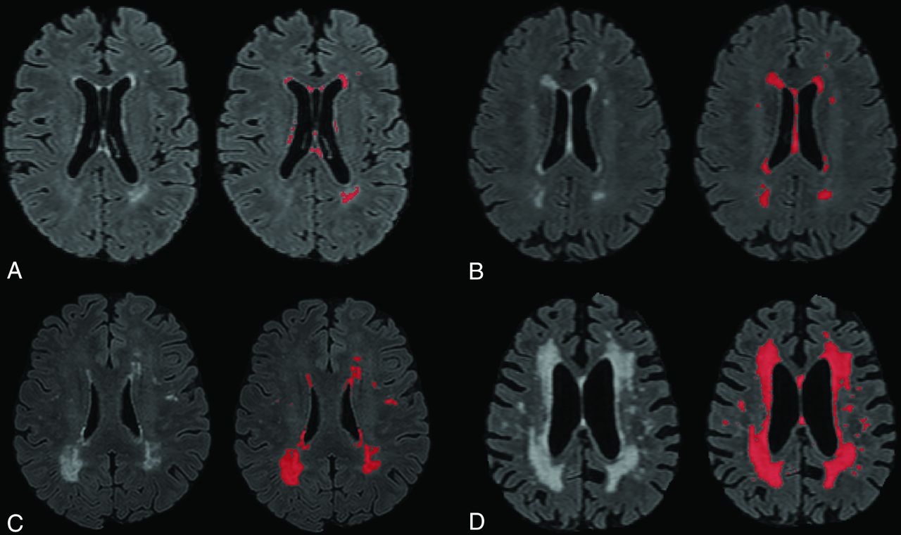
- Fig 1.
Four sample images of WMH on T2 FLAIR, labeled for quantification using an intensity threshold with our software. A, Minimal WMHs. B, Mild WMHs. C, Moderate WMHs. D, Severe WMHs.
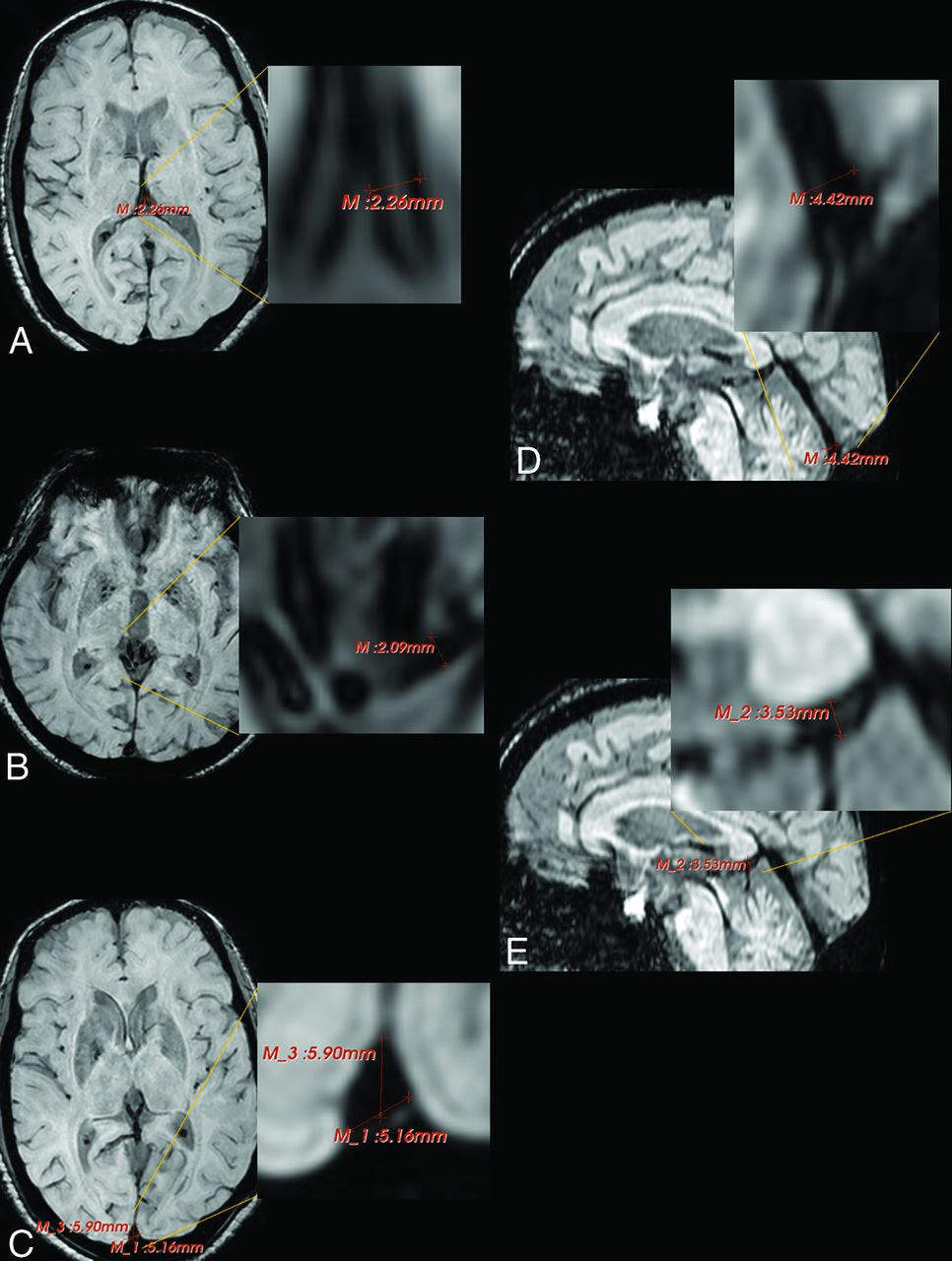
- Fig 2.
Sample depiction of vein/sinus measurement on SWI using 3D Slicer. A, The diameters of the left and right internal cerebral veins were measured in the axial plane and then averaged. B, The diameters of the left and right basal veins of Rosenthal were measured at their termini in the axial plane and then averaged. C, The base and anterior-posterior diameters of the superior sagittal plane were measured in the axial plane and then averaged. D, The straight sinus terminus was measured in the sagittal plane. E, The vein of Galen was measured inferior to the splenium in the sagittal plane.

Tables
- Table 1:
Study sample characteristics and mean values for measured vein/sinus diameters and WMH volume
Characteristics No., overall 682 Age at scan (mean) (SD) (yr) 73.9 (5.93) Women (No.) (% total) 403 (59.1) Race/ethnicity (No.) (% within race/ethnicity) White 185 (27.1) Black 243 (35.6) Hispanic 236 (34.6) Other 18 (2.6) Vascular risk factors (No.) (% total) Heart disease 130 (19.1) Hypertension 434 (63.6) Diabetes 166 (24.3) Diameter, based on SWI (mean) (SD (mm) Internal cerebral veins 1.73 (0.26) Basal veins of Rosenthal 1.64 (0.26) Superior sagittal sinus 6.18 (0.87) Vein of Galen 2.72 (0.57) Straight sinus 3.80 (0.90) WMH volume (mean) (SD) (mm3) Total 5.21 (6.64) Frontal lobe 2.27 (3.46) Temporal lobe 0.32 (0.57) Parietal lobe 1.44 (2.46) Occipital lobe 0.42 (0.56) - Table 2:
Unadjusted associations (using standardized β values and corresponding 95% CI) between sinus/vein diameters and WMH volumea
Total WMH Frontal Lobe Temporal Lobe Parietal Lobe Occipital Lobe Internal cerebral veins β (95% CI) 0.11 (0.03–0.18) 0.10 (0.03–0.18) 0.11 (0.03–0.18) 0.12 (0.05–0.20) 0.09 (0.02–0.17) P .006 .007 .006 .001 .02 Basal veins of Rosenthal β (95% CI) 0.13 (0.06–0.20) 0.15 (0.08–0.22) 0.13 (0.05–0.20) 0.10 (0.02–0.17) 0.06 (−0.01–0.13) P .001 <.001 .001 .009 .11 Superior sagittal sinus β (95% CI) 0.09 (0.01–0.16) 0.07 (−0.01–0.14) 0.12 (0.05–0.20) 0.09 (0.02–0.17) 0.11 (0.04–0.19) P .02 .08 .002 .02 .003 Vein of Galen β (95% CI) 0.02 (−0.05–0.10) 0.05 (−0.03–0.12) -0.01 (−0.08–0.07) 0.01 (−0.06–0.09) 0.00 (−0.08–0.07) P .55 .25 .84 .72 .95 Straight sinus β (95% CI) 0.00 (−0.08–0.07) 0.00 (−0.08–0.07) -0.01 (−0.08–0.07) 0.00 (−0.07–0.08) 0.00 (−0.08–0.07) P .98 .97 .88 .96 .93 ↵a Model 1: unadjusted; model 2: age-adjusted; model 3: adjusted for age and estimated intracranial volume.
- Table 3:
Associations (using standardized β values and corresponding 95% CI) between sinus/vein diameters and WMH volume after adjusting for age, estimated intracranial volume, and vascular risk factors
Total WMH Frontal Lobe Temporal Lobe Parietal Lobe Occipital Lobe Internal cerebral veins β (95% CI) 0.09 (0.02–0.16) 0.09 (0.01–0.16) 0.09 (0.02–0.17) 0.10 (0.03–0.18) 0.07 (−0.01–0.14) P .02 .02 .02 .006 .09 Basal veins of Rosenthal β (95% CI) 0.10 (0.02–0.17) 0.11 (0.04–0.19) 0.09 (0.01–0.17) 0.07 (−0.01–0.14) 0.00 (−0.07–0.08) P .01 .003 .02 .09 .95 Superior sagittal sinus β (95% CI) 0.04 (−0.4–0.12) 0.01 (−0.07–0.09) 0.08 (−0.001–0.16) 0.05 (−0.03–0.12) 0.04 (−0.04–0.12) P .33 .75 .05 .26 .35 Vein of Galen β (95% CI) 0.01 (−0.07–0.08) 0.03 (−0.05–0.10) −0.02 (−0.10–0.06) 0.00 (−0.07–0.08) −0.02 (−0.10–0.05) P .85 .51 .61 .93 .54 Straight sinus β (95% CI) -0.01 (−0.08–0.07) -0.01 (−0.08–0.06) −0.01 (−0.09–0.06) -0.003 (−0.08–0.07) -0.02 (−0.10–0.05) P .83 .79 .71 .93 .55


