
Abstract
SUMMARY: In this retrospective case-control study, we investigated whether an image-processing algorithm designed to exaggerate the intensity of diseased hippocampi on FLAIR images can improve the diagnostic accuracy and interobserver reliability of radiologists in detecting mesial temporal sclerosis–related hippocampal signal alteration. Herein, we share the results of this study that showed that the image processing improved the confidence of radiologists in detecting mesial temporal sclerosis–related signal alteration, allowing an improved sensitivity, specificity, and interobserver reliability.
ABBREVIATIONS:
- MTS
- mesial temporal sclerosis
- SI
- signal intensity
A confident identification of mesial temporal sclerosis (MTS) based on hippocampal volume loss, signal abnormality, and architectural distortion on MR imaging is important for timely surgical management of refractory epilepsy.1⇓⇓–4 Because signal abnormalities may precede volume loss in MTS, improved detection of hippocampal signal abnormalities may help in its early diagnosis and treatment.5 Recently, an image-processing algorithm using correlative properties of neighboring pixels has shown promise in selectively enhancing the visual conspicuity of diseased hippocampi.6,7 Here we share the results of a retrospective case-control study testing whether this image processing could translate into an improved diagnostic performance of radiologists in detecting MTS in adults.
Materials and Methods
Image Processing and Review
A blinded coinvestigator processed coronal FLAIR images from 51 subjects with MTS and 51 healthy controls (Table 1) with a proprietary image-processing algorithm (Correlative Image Enhancement) using a custom plug-in for OsiriX Lite (https://www.downloadthat.com/windows/search/osirix_lite_for_windows). The algorithm exaggerated the hippocampal signal intensity (SI) if it was considerably higher than that of the normal gray matter as defined by an ROI drawn over the normal-appearing insular cortex. Processed images were saved as a separate DICOM series. Six readers with varied experience levels reviewed baseline and processed images separately, rating the SI of each hippocampus on a 5-point scale: 1, definitely normal; 2, probably normal; 3, possibly normal; 4, probably abnormal; and 5, definitely abnormal. They also indicated whether the hippocampal SI was unusually high, suggesting the effect of processing.
Details of cases and controls
Data Analysis
Differences among the median SI ratings across all readers for baseline and processed images were computed and compared using a paired t test or signed rank test. If one considered the SI ratings of 1–3 as normal and 4–5 as abnormal, the effects of processing on the sensitivity, specificity, positive predictive value, negative predictive value, and accuracy of each reader and for all readers were assessed. Sensitivity was additionally assessed for a subgroup of patients in whom the MR imaging findings were originally reported as normal. The effect of processing on the interobserver reliability of identifying hippocampal signal abnormality (SI ratings) was computed using a model-based measure of agreement, which is robust to the underlying disease prevalence.8
Results
All readers reported confluent areas of markedly increased signal (Fig 1) in processed images of 37 (72.5%) diseased hippocampi. Individual readers reported processing-related foci of markedly increased SI in 2%–4.9% (average, 3.6%) of control hippocampi (Fig 2).

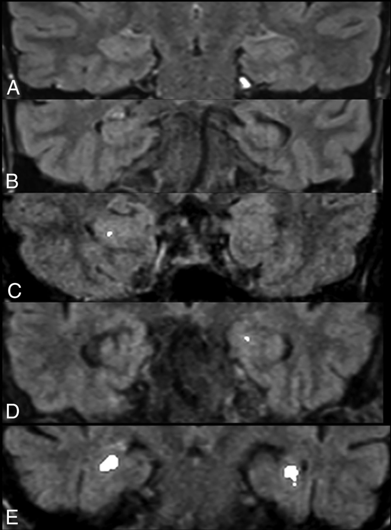
Coronal FLAIR images (A–E) across the medial temporal lobes in patients with pathologically confirmed right-sided (A, C, and E) and left-sided (B and D) MTS. Corresponding images following image processing (A1–E1) demonstrate confluent areas of marked exaggeration of signal intensity of the diseased hippocampi. Note a similar exaggeration of signal intensity to a smaller extent in the right hippocampus (arrow) in B1, presumably indicating bilateral disease.
Representative postprocessed coronal FLAIR images across the medial temporal lobes of 5 controls without seizures. Processing did not result in any alteration of hippocampal signal (A and B) for most control hippocampi. While punctate (C and D) foci of signal exaggeration were noted in some control hippocampi, false-positive confluent regions of increased intensity mimicking MTS (E) were observed in the bilateral hippocampi of 1 (2%) control.
Effect on Confidence Ratings
Image processing resulted in an increase in median SI rating for diseased hippocampi (P < .001, Table 2) and a decrease in the median SI rating for both left- (P = .03) and right-sided (P = .003) hippocampi in controls.
Effect of image processing on signal intensity ratings for subjects with MTS, right-sided control hippocampi, and left-sided control hippocampi on a 5-point scalea
Effect on Readers' Diagnostic Performance
An improvement in the average sensitivity, specificity, positive predictive value, negative predictive value, and accuracy was seen after processing (Table 3). The average sensitivity increased by >10% (Table 3), and readers were able to detect hippocampal signal abnormality in 4/10 (40%) cases reported as having normal findings on the original clinical reports.
Diagnostic performance of 6 blinded readers in detection of MTS-related hippocampal signal alteration before and after image processing with a proprietary algorithm
Effect on Interobserver Reliability
Processing resulted in an improvement in the interobserver agreement in SI ratings for cases of MTS from moderate (Fleiss κ = 0.4; 95% CI, 0.2–0.56) to almost perfect (Fleiss κ = 0.88; 95% CI, 0.80–0.97).9 For control hippocampi, interobserver agreement improved from slight to moderate.9
Discussion
In our study, an easily recognizable increase in the contrast-to-noise ratio of diseased hippocampi by the image processing6,7 translated into substantial improvement in sensitivity when present and slight improvement in specificity when absent. Our results indicate that this processing may help detect MTS in some patients with focal epilepsy with otherwise normal MR imaging findings,10 thereby allowing earlier diagnosis and treatment. An improvement in interobserver agreement may make this processing particularly helpful for nonexpert readers, prompting a timely referral to experts.
Unlike volumetry-based techniques,11,12 the image processing used in our study relies on highlighting MTS-related signal abnormality. Thus, it is similar to other techniques such as T2 relaxometry and automated FLAIR analysis but may be more generalizable because it does not require comparison with normative data.13⇓⇓–16
Conclusions
The image-processing algorithm tested by us can improve detection of MTS on MR imaging. In view of a small possibility of false-positive effects of processing, this technique should serve as a complement to a complete epilepsy protocol MR imaging, interpreted in the context of clinical history.
Footnotes
Disclosures: Amber Salter—UNRELATED: Consultancy: Circulation Cardiovascular Imaging. Aseem Sharma—RELATED: Other: Correlative Enhancement LLC, Comments: I hold the intellectual property rights to the image-processing algorithms used in this study. I have founded a company (Correlative Enhancement LLC) with the aim of future commercialization of this intellectual property. I am the sole proprietor of the company, and since the inception of the company until now (including the time during which I processed the images for this study), this company has not received funding from any external source. While I used the algorithms to process the images for this study, I did not participate in patient selection, image review, image analysis, or the subsequent statistical analysis; UNRELATED: Consultancy: Biomedical Systems, Comments: As a consultant, I serve as an independent reviewer for imaging studies performed for research by third parties; Patents (Planned, Pending or Issued): I have been issued the patent for the method of image processing used in this and other studies; Stock/Stock Options: GE Healthcare, Comments: I hold approximately $10,000 worth of publicly traded GE stocks. Sonika Dahiya—UNRELATED: Employment: Washington University School of Medicine.
REFERENCES
- Received October 26, 2018.
- Accepted after revision January 18, 2019.
- © 2019 by American Journal of Neuroradiology


Jump to section
Related Articles
Cited By...
- No citing articles found.