Advertisement

Main menu

  • Home
  • Content
    • Current Issue
    • Accepted Manuscripts
    • Article Preview
    • Past Issue Archive
    • Video Articles
    • AJNR Case Collection
    • Case of the Week Archive
    • Case of the Month Archive
    • Classic Case Archive
  • Special Collections
    • AJNR Awards
    • ASNR Foundation Special Collection
    • Most Impactful AJNR Articles
    • Photon-Counting CT
    • Spinal CSF Leak Articles (Jan 2020-June 2024)
  • Multimedia
    • AJNR Podcasts
    • AJNR SCANtastic
    • Trainee Corner
    • MRI Safety Corner
    • Imaging Protocols
  • For Authors
    • Submit a Manuscript
    • Submit a Video Article
    • Submit an eLetter to the Editor/Response
    • Manuscript Submission Guidelines
    • Statistical Tips
    • Fast Publishing of Accepted Manuscripts
    • Graphical Abstract Preparation
    • Imaging Protocol Submission
    • Author Policies
  • About Us
    • About AJNR
    • Editorial Board
    • Editorial Board Alumni
  • More
    • Become a Reviewer/Academy of Reviewers
    • Subscribers
    • Permissions
    • Alerts
    • Feedback
    • Advertisers
    • ASNR Home

User menu

  • Alerts
  • Log in

Search

  • Advanced search
American Journal of Neuroradiology

American Journal of Neuroradiology

ASHNR American Society of Functional Neuroradiology ASHNR American Society of Pediatric Neuroradiology ASSR
  • Alerts
  • Log in

Advanced Search

  • Home
  • Content
    • Current Issue
    • Accepted Manuscripts
    • Article Preview
    • Past Issue Archive
    • Video Articles
    • AJNR Case Collection
    • Case of the Week Archive
    • Case of the Month Archive
    • Classic Case Archive
  • Special Collections
    • AJNR Awards
    • ASNR Foundation Special Collection
    • Most Impactful AJNR Articles
    • Photon-Counting CT
    • Spinal CSF Leak Articles (Jan 2020-June 2024)
  • Multimedia
    • AJNR Podcasts
    • AJNR SCANtastic
    • Trainee Corner
    • MRI Safety Corner
    • Imaging Protocols
  • For Authors
    • Submit a Manuscript
    • Submit a Video Article
    • Submit an eLetter to the Editor/Response
    • Manuscript Submission Guidelines
    • Statistical Tips
    • Fast Publishing of Accepted Manuscripts
    • Graphical Abstract Preparation
    • Imaging Protocol Submission
    • Author Policies
  • About Us
    • About AJNR
    • Editorial Board
    • Editorial Board Alumni
  • More
    • Become a Reviewer/Academy of Reviewers
    • Subscribers
    • Permissions
    • Alerts
    • Feedback
    • Advertisers
    • ASNR Home
  • Follow AJNR on Twitter
  • Visit AJNR on Facebook
  • Follow AJNR on Instagram
  • Join AJNR on LinkedIn
  • RSS Feeds

AJNR is seeking candidates for the AJNR Podcast Editor. Read the position description.

Research ArticleAdult Brain

Quantification of Oscillatory Shear Stress from Reciprocating CSF Motion on 4D Flow Imaging

S. Yamada, H. Ito, M. Ishikawa, K. Yamamoto, M. Yamaguchi, M. Oshima and K. Nozaki
American Journal of Neuroradiology March 2021, 42 (3) 479-486; DOI: https://doi.org/10.3174/ajnr.A6941
S. Yamada
aFrom the Department of Neurosurgery (S.Y., K.N.), Shiga University of Medical Science, Shiga, Japan
bDepartment of Neurosurgery and Normal Pressure Hydrocephalus Center (S.Y., K.Y., M.Y.), Rakuwakai Otowa Hospital, Kyoto, Japan
cInterfaculty Initiative in Information Studies/Institute of Industrial Science (S.Y., M.O.), The University of Tokyo, Tokyo, Japan
  • Find this author on Google Scholar
  • Find this author on PubMed
  • Search for this author on this site
  • ORCID record for S. Yamada
H. Ito
dMedical System Research and Development Center (H.I.), Fujifilm Corporation, Tokyo, Japan
  • Find this author on Google Scholar
  • Find this author on PubMed
  • Search for this author on this site
  • ORCID record for H. Ito
M. Ishikawa
eRakuwa Villa Ilios (M.I.), Kyoto, Japan
  • Find this author on Google Scholar
  • Find this author on PubMed
  • Search for this author on this site
  • ORCID record for M. Ishikawa
K. Yamamoto
bDepartment of Neurosurgery and Normal Pressure Hydrocephalus Center (S.Y., K.Y., M.Y.), Rakuwakai Otowa Hospital, Kyoto, Japan
  • Find this author on Google Scholar
  • Find this author on PubMed
  • Search for this author on this site
  • ORCID record for K. Yamamoto
M. Yamaguchi
bDepartment of Neurosurgery and Normal Pressure Hydrocephalus Center (S.Y., K.Y., M.Y.), Rakuwakai Otowa Hospital, Kyoto, Japan
  • Find this author on Google Scholar
  • Find this author on PubMed
  • Search for this author on this site
  • ORCID record for M. Yamaguchi
M. Oshima
cInterfaculty Initiative in Information Studies/Institute of Industrial Science (S.Y., M.O.), The University of Tokyo, Tokyo, Japan
  • Find this author on Google Scholar
  • Find this author on PubMed
  • Search for this author on this site
  • ORCID record for M. Oshima
K. Nozaki
aFrom the Department of Neurosurgery (S.Y., K.N.), Shiga University of Medical Science, Shiga, Japan
  • Find this author on Google Scholar
  • Find this author on PubMed
  • Search for this author on this site
  • ORCID record for K. Nozaki
  • Article
  • Figures & Data
  • Supplemental
  • Info & Metrics
  • Responses
  • References
  • PDF
Loading

Article Figures & Data

Figures

  • Tables
  • FIG 1.
    • Download figure
    • Open in new tab
    • Download powerpoint
    FIG 1.

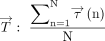
    Schema explaining OSS: θ(n) is the angle between Embedded Image and Embedded Image in the nth phase Embedded Image(n) Embedded Image Where Embedded Image indicates a vector of TAWSS, and Embedded Image indicates a vector of WSS.

  • FIG 2.
    • Download figure
    • Open in new tab
    • Download powerpoint
    FIG 2.

    Oscillation of WSS due to bidirectional CSF flow through the cerebral aqueduct. The numbers 1, 2, and 3 demonstrate temporal changes of the reciprocating CSF motion during 1 cardiac cycle in a 77-year-old man diagnosed with iNPH. The colored path lines (lower figures) show the flow as a moving trajectory of the virtual particle through the middle part of the cerebral aqueduct in 1 heartbeat. The color and length of 3D streamlines (upper figures) indicate the flow velocity as pink for fast and blue for slow. The sampling interval of streamlines and path lines was set at 0.2 mm after the trilinear interpolation. The arrows indicate the WSS vector produced by the flow parallel to the wall surface. The size and direction of the arrows indicate the magnitude and direction of the WSS vector. Temporal changes of the WSS vector at the dorsal region (yellow arrows) were larger than those at the ventral region (white arrows).

  • FIG 3.
    • Download figure
    • Open in new tab
    • Download powerpoint
    FIG 3.

    Distributions and fluctuations of shear stress parameters per heartbeat in iNPH. The colored surfaces in the ventricles show the distribution of WSS magnitude (A) and OSI (B) in the same patient with iNPH, as shown in Fig 2. Red indicates high and blue indicates low. Line-graphs C and D show chronologic changes of WSS magnitude and OSS at the dorsal region of the foramen magnum (ROI 2, yellow) and dorsal (ROI 3, red) and ventral (ROI 4, blue) regions of the cerebral aqueduct in 1 cardiac cycle. Considering the shear stress direction, these changes were completely different.

  • FIG 4.
    • Download figure
    • Open in new tab
    • Download powerpoint
    FIG 4.

    Boxplots for the amplitude of OSS and TAWSS at the dorsal and ventral regions of the cerebral aqueduct among 3 groups. A, Distribution of the OSS amplitude at the dorsal region of the cerebral aqueduct. B, Distribution of the TAWSS at the dorsal region of the cerebral aqueduct. C, Distribution of the OSS amplitude at the ventral region of the cerebral aqueduct. D, Distribution of the TAWSS at the ventral region of the cerebral aqueduct.

Tables

  • Figures
    • View popup
    Table 1:

    Clinical and morphologic characteristics of the study populationa

    iNPH OnlyiNPH with ADControlP Value
    Total number41239
    Male/female28:1313:104:5.332
    Age (yr)78.2 [SD, 6.62]79.8 [SD, 5.83]75.7 [SD, 5.70].207
    Anteroposterior diameter (mm)
     Foramen of Magendie5.72 [SD, 1.77]5.88 [SD, 2.04]2.02 [SD, 0.44]<.001
     Lower end of cerebral aqueduct4.44 [SD, 0.87]4.25 [SD, 1.41]2.23 [SD, 0.70]<.001
     Upper end of cerebral aqueduct3.65 [SD, 0.66]3.45 [SD, 0.73]2.30 [SD, 0.54]<.001
     Midbrain8.56 [SD, 0.88]8.43 [SD, 0.92]9.32 [SD, 1.57].230
    Stroke volume (μL/heartbeat)
     Foramen of Magendie32.9 [SD, 22.6]37.4 [SD, 37.6]21.1 [SD, 11.7].448
     Lower end of cerebral aqueduct37.6 [SD, 28.9]39.6 [SD, 43.7]17.8 [SD, 13.0].083
     Upper end of cerebral aqueduct49.0 [SD, 33.3]52.6 [SD, 32.5]18.9 [SD, 10.5].002
     Foramina of Monro96.5 [SD, 41.0]97.8 [SD, 39.1]62.8 [SD, 44.6].042
    • a Data are means. P value is from the Kruskal-Wallis rank sum test.

    • View popup
    Table 2:

    Mean value [SD] of parameters at the cerebral aqueduct in the study populationa

    All (n = 73)iNPH Only (n = 41)iNPH with AD (n = 23)Control (n = 9)P1P2P3
    Dorsal region of the cerebral aqueduct
     OSS amplitude52.5 [SD, 33.9]59.7 [SD, 34.1]52.9 [SD, 32.2]19.8 [SD, 13.3].483<.001.004
     Maximum OSS35.9 [SD, 16.0]38.7 [SD, 15.5]37.3 [SD, 16.1]19.7 [SD, 6.3].566<.001.002
     Minimum OSS–16.7 [SD, 19.8]–21.0 [SD, 20.2]–15.6 [SD, 18.9]–0.1 [SD, 9.6].277.001.043
     TAWSS18.2 [SD, 8.9]19.7 [SD, 8.7]18.8 [SD, 9.3]9.9 [SD, 2.2].678<.001.008
     OSI0.21 [SD, 0.13]0.24 [SD, 0.12]0.19 [SD, 0.14]0.16 [SD, 0.11].234.077.614
    Ventral region of the cerebral aqueduct
     OSS amplitude27.2 [SD, 28.4]36.0 [SD, 32.2]18.1 [SD, 19.8]11.9 [SD, 11.1].022.020.363
     Maximum OSS23.6 [SD, 15.9]28.8 [SD, 17.9]18.2 [SD, 10.9]14.2 [SD, 6.4].017.011.246
     Minimum OSS–3.6 [SD, 13.9]–7.1 [SD, 15.9]0.14 [SD, 10.1]2.3 [SD, 7.5].080.091.592
     TAWSS12.2 [SD, 7.1]14.3 [SD, 8.1]10.1 [SD, 4.9]8.1 [SD, 2.7].038.020.458
     OSI0.13 [SD, 0.15]0.15 [SD, 0.15]0.10 [SD, 0.14]0.11 [SD, 0.15].114.355.910
    • ↵a P1 indicates probability value of iNPH only versus iNPH with AD by the Wilcoxon rank sum test; P2, probability value of iNPH only versus controls by the Wilcoxon rank sum test; and P3, probability value of iNPH with AD versus controls by the Wilcoxon rank sum test.

    • View popup
    Table 3:

    Relationships with anterior-posterior diameter by the Pearson correlation coefficient (95% confidence intervals)

    Foramen of MagendieLower End of Cerebral AqueductUpper End of Cerebral AqueductMidbrain
    Dorsal region of the cerebral aqueduct
     OSS amplitude0.55 (0.36–0.69)0.47 (0.26–0.63)0.28 (0.05–0.48)NS
     Maximum OSS0.53 (0.34–0.68)0.41 (0.20–0.59)NSNS
     Minimum OSS–0.51 (–0.66 to –0.31)–0.46 (–0.63 to –0.26)–0.32 (–0.51 to –0.10)NS
     TAWSS0.56 (0.37–0.70)0.40 (0.18–0.58)NSNS
     OSINSNSNSNS
    Ventral region of the cerebral aqueduct
     OSS amplitudeNS0.27 (0.04–0.47)NSNS
     Maximum OSSNS0.31 (0.08–0.50)NSNS
     Minimum OSSNSNSNSNS
     TAWSS0.26 (0.04–0.47)0.33 (0.10–0.52)NSNS
     OSINSNSNSNS
    • Note:—NS indicates not significant (P ≥ .05) by the Pearson correlation analysis.

    • View popup
    Table 4:

    Relationships with stroke volume by the Pearson correlation coefficient (95% confidence intervals)

    Foramen of MagendieLower End of Cerebral AqueductUpper End of Cerebral AqueductForamina of Monro
    Dorsal region of the cerebral aqueduct
     OSS amplitude0.48 (0.28–0.64)0.53 (0.34–0.68)0.63 (0.47–0.76)NS
     Maximum OSS0.50 (0.31–0.66)0.56 (0.37–0.70)0.66 (0.50–0.77)NS
     Minimum OSS–0.41 (–0.59 to –0.20)–0.46 (–0.63 to –0.26)–0.55 (–0.70 to –0.37)NS
     TAWSS0.43 (0.22–0.60)0.53 (0.34–0.68)0.73 (0.60–0.82)NS
     OSINSNSNSNS
    Ventral region of the cerebral aqueduct
     OSS amplitudeNS0.30 (0.07–0.50)0.25 (0.02–0.46)NS
     Maximum OSSNS0.32 (0.10–0.52)0.29 (0.07–0.49)NS
     Minimum OSSNS–0.24 (–0.45 to –0.01)NSNS
     TAWSSNS0.37 (0.15–0.55)0.34 (0.12–0.53)NS
     OSINSNSNSNS
    • Note:—NS indicates not significant (P ≥ .05) by the Pearson correlation analysis.

PreviousNext
Back to top

In this issue

American Journal of Neuroradiology: 42 (3)
American Journal of Neuroradiology
Vol. 42, Issue 3
1 Mar 2021
  • Table of Contents
  • Index by author
  • Complete Issue (PDF)
Advertisement
Print
Download PDF
Email Article

Thank you for your interest in spreading the word on American Journal of Neuroradiology.

NOTE: We only request your email address so that the person you are recommending the page to knows that you wanted them to see it, and that it is not junk mail. We do not capture any email address.

Enter multiple addresses on separate lines or separate them with commas.
Quantification of Oscillatory Shear Stress from Reciprocating CSF Motion on 4D Flow Imaging
(Your Name) has sent you a message from American Journal of Neuroradiology
(Your Name) thought you would like to see the American Journal of Neuroradiology web site.
CAPTCHA
This question is for testing whether or not you are a human visitor and to prevent automated spam submissions.
Cite this article
S. Yamada, H. Ito, M. Ishikawa, K. Yamamoto, M. Yamaguchi, M. Oshima, K. Nozaki
Quantification of Oscillatory Shear Stress from Reciprocating CSF Motion on 4D Flow Imaging
American Journal of Neuroradiology Mar 2021, 42 (3) 479-486; DOI: 10.3174/ajnr.A6941

Citation Manager Formats

  • BibTeX
  • Bookends
  • EasyBib
  • EndNote (tagged)
  • EndNote 8 (xml)
  • Medlars
  • Mendeley
  • Papers
  • RefWorks Tagged
  • Ref Manager
  • RIS
  • Zotero
0 Responses
Respond to this article
Share
Bookmark this article
Quantification of Oscillatory Shear Stress from Reciprocating CSF Motion on 4D Flow Imaging
S. Yamada, H. Ito, M. Ishikawa, K. Yamamoto, M. Yamaguchi, M. Oshima, K. Nozaki
American Journal of Neuroradiology Mar 2021, 42 (3) 479-486; DOI: 10.3174/ajnr.A6941
del.icio.us logo Twitter logo Facebook logo Mendeley logo
  • Tweet Widget
  • Facebook Like
  • Google Plus One
Purchase

Jump to section

  • Article
    • Abstract
    • ABBREVIATIONS:
    • MATERIALS AND METHODS
    • RESULTS
    • DISCUSSION
    • CONCLUSIONS
    • Acknowledgments
    • Footnotes
    • References
  • Figures & Data
  • Supplemental
  • Info & Metrics
  • Responses
  • References
  • PDF

Related Articles

  • PubMed
  • Google Scholar

Cited By...

  • No citing articles found.
  • Crossref (19)
  • Google Scholar

This article has been cited by the following articles in journals that are participating in Crossref Cited-by Linking.

  • Exploring mechanisms of ventricular enlargement in idiopathic normal pressure hydrocephalus: a role of cerebrospinal fluid dynamics and motile cilia
    Shigeki Yamada, Masatsune Ishikawa, Kazuhiko Nozaki
    Fluids and Barriers of the CNS 2021 18 1
  • Aging-related volume changes in the brain and cerebrospinal fluid using artificial intelligence-automated segmentation
    Shigeki Yamada, Tomohiro Otani, Satoshi Ii, Hiroto Kawano, Kazuhiko Nozaki, Shigeo Wada, Marie Oshima, Yoshiyuki Watanabe
    European Radiology 2023 33 10
  • Four‐dimensional flow MRI for quantitative assessment of cerebrospinal fluid dynamics: Status and opportunities
    Leonardo A. Rivera‐Rivera, Tomas Vikner, Laura Eisenmenger, Sterling C. Johnson, Kevin M. Johnson
    NMR in Biomedicine 2024 37 7
  • Diffusion magnetic resonance imaging of cerebrospinal fluid dynamics: Current techniques and future advancements
    Adam M. Wright, Yu‐Chien Wu, Li Feng, Qiuting Wen
    NMR in Biomedicine 2024 37 9
  • Usefulness of intravoxel incoherent motion MRI for visualizing slow cerebrospinal fluid motion
    Shigeki Yamada, Shinnosuke Hiratsuka, Tomohiro Otani, Satoshi Ii, Shigeo Wada, Marie Oshima, Kazuhiko Nozaki, Yoshiyuki Watanabe
    Fluids and Barriers of the CNS 2023 20 1
  • A subject‐specific assessment of measurement errors and their correction in cerebrospinal fluid velocity maps using 4D flow MRI
    Selin Yavuz Ilik, Tomohiro Otani, Shigeki Yamada, Yoshiyuki Watanabe, Shigeo Wada
    Magnetic Resonance in Medicine 2022 87 5
  • Sustained glymphatic transport and impaired drainage to the nasal cavity observed in multiciliated cell ciliopathies with hydrocephalus
    Yuechuan Xue, Zachary Gursky, Brittany Monte, Sunil Koundal, Xiaodan Liu, Hedok Lee, Tatyana V. Michurina, Kennelia A. Mellanson, Lucy Zhao, Alice Nemajerova, Kristopher T. Kahle, Ken-Ichi Takemaru, Grigori Enikolopov, Natalia I. Peunova, Helene Benveniste
    Fluids and Barriers of the CNS 2022 19 1
  • Age-Related Changes in Cerebrospinal Fluid Dynamics in the Pathogenesis of Chronic Hydrocephalus in Adults
    Shigeki Yamada, Hirotaka Ito, Motoki Tanikawa, Satoshi Ii, Tomohiro Otani, Shigeo Wada, Marie Oshima, Yoshiyuki Watanabe, Mitsuhito Mase
    World Neurosurgery 2023 178
  • Automatic assessment of disproportionately enlarged subarachnoid-space hydrocephalus from 3D MRI using two deep learning models
    Shigeki Yamada, Hirotaka Ito, Hironori Matsumasa, Satoshi Ii, Tomohiro Otani, Motoki Tanikawa, Chifumi Iseki, Yoshiyuki Watanabe, Shigeo Wada, Marie Oshima, Mitsuhito Mase
    Frontiers in Aging Neuroscience 2024 16
  • Biomechanical effects of hyper-dynamic cerebrospinal fluid flow through the cerebral aqueduct in idiopathic normal pressure hydrocephalus patients
    Shusaku Maeda, Tomohiro Otani, Shigeki Yamada, Yoshiyuki Watanabe, Selin Yavuz Ilik, Shigeo Wada
    Journal of Biomechanics 2023 156

More in this TOC Section

  • Diagnostic Neuroradiology of Monoclonal Antibodies
  • Clinical Outcomes After Chiari I Decompression
  • Segmentation of Brain Metastases with BLAST
Show more Adult Brain

Similar Articles

Advertisement

Indexed Content

  • Current Issue
  • Accepted Manuscripts
  • Article Preview
  • Past Issues
  • Editorials
  • Editor's Choice
  • Fellows' Journal Club
  • Letters to the Editor
  • Video Articles

Cases

  • Case Collection
  • Archive - Case of the Week
  • Archive - Case of the Month
  • Archive - Classic Case

Special Collections

  • AJNR Awards
  • ASNR Foundation Special Collection
  • Most Impactful AJNR Articles
  • Photon-Counting CT
  • Spinal CSF Leak Articles (Jan 2020-June 2024)

More from AJNR

  • Trainee Corner
  • Imaging Protocols
  • MRI Safety Corner

Multimedia

  • AJNR Podcasts
  • AJNR Scantastics

Resources

  • Turnaround Time
  • Submit a Manuscript
  • Submit a Video Article
  • Submit an eLetter to the Editor/Response
  • Manuscript Submission Guidelines
  • Statistical Tips
  • Fast Publishing of Accepted Manuscripts
  • Graphical Abstract Preparation
  • Imaging Protocol Submission
  • Evidence-Based Medicine Level Guide
  • Publishing Checklists
  • Author Policies
  • Become a Reviewer/Academy of Reviewers
  • News and Updates

About Us

  • About AJNR
  • Editorial Board
  • Editorial Board Alumni
  • Alerts
  • Permissions
  • Not an AJNR Subscriber? Join Now
  • Advertise with Us
  • Librarian Resources
  • Feedback
  • Terms and Conditions
  • AJNR Editorial Board Alumni

American Society of Neuroradiology

  • Not an ASNR Member? Join Now

© 2025 by the American Society of Neuroradiology All rights, including for text and data mining, AI training, and similar technologies, are reserved.
Print ISSN: 0195-6108 Online ISSN: 1936-959X

Powered by HighWire