
Article Figures & Data
Figures
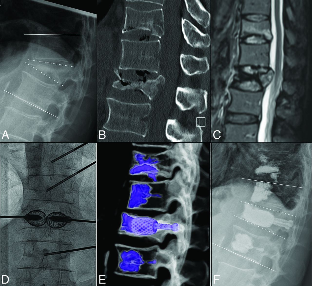
- FIG 1.
A, Procedural steps of the SAIF technique. Preprocedural lateral view of a T11 VP fracture. B, Balloon-mounted vertebral body stent insertion in the vertebral body. C, Balloon expansion of the stents. D, Access trocars are exchanged with transpedicular, cannulated-fenestrated screws over a Kirschner wire. Anterior-posterior and lateral views (E and F) obtained before cement injection through the screws.
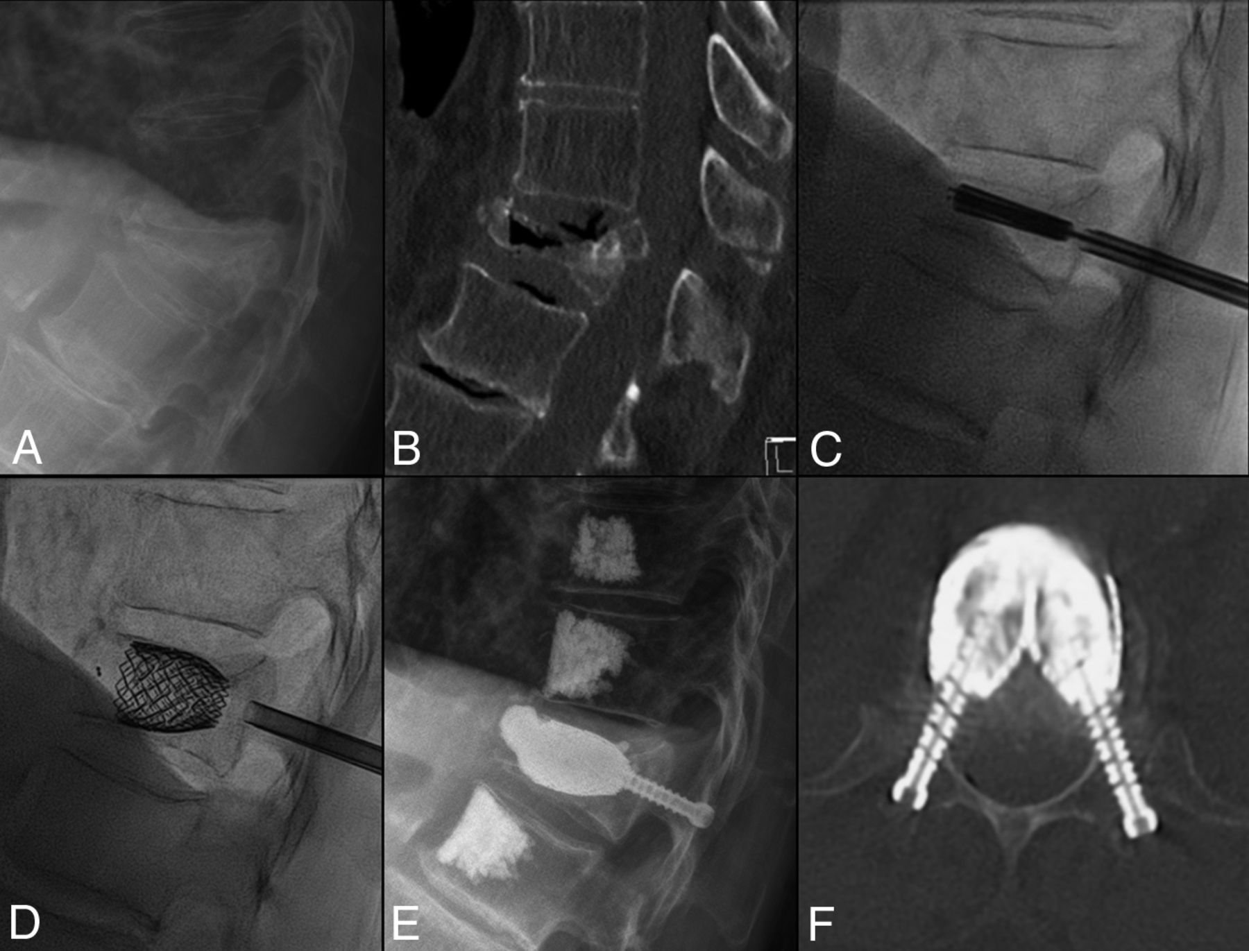
- FIG 2.
A, Standing plain film shows a L1 VP with kyphotic angulation. White lines along the endplates of T12 and L2 indicate the LKA, while the dashed white lines along the L1 endplates indicate the VKA. B, Sagittal CT shows a pseudoarthrosis with a gas cleft in L1 and increased vertebral body height in supine decubitus positioning, in keeping with a mobile fracture. An additional fracture of T11 was treated with vertebroplasty. Sagittal fat-suppressed T2WI (C) shows posterior wall retropulsion and central canal stenosis without cord compression and an additional milder fracture at T11. Anterior-posterior intraprocedural fluoroscopic image (D) demonstrates SAIF implants, with pedicular screws inserted in the expanded stents before cement injection. Volume-rendering postprocedure CT (E) shows the SAIF treatment of L1 and vertebral augmentation at T11, T12, and L2. Postprocedural standing plain film (F) shows reduction of the LKA from 28° to 16° and of the VKA from 30° to 11°.
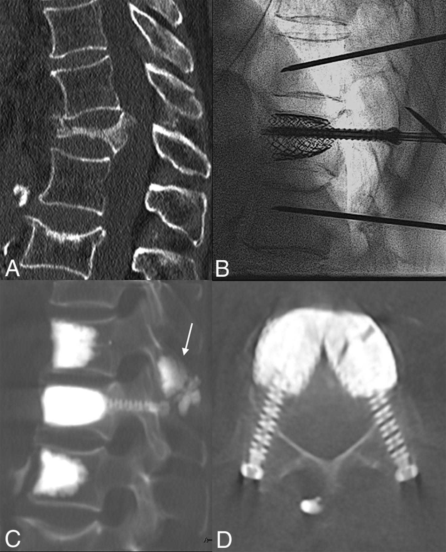
- FIG 3.
Sagittal CT (A) shows a T12 VP, with segmental kyphosis and a T11 spinous process fracture. Intraprocedural fluoroscopic lateral view (B) shows fracture reduction by the SAIF technique before cement augmentation. Postprocedural sagittal (C) and axial (D) CT images show the final results obtained with the SAIF construct. There is cement augmentation of the T11 spinous process fracture (arrow), which was particularly tender at palpation, and the prophylactic augmentation of the adjacent levels.
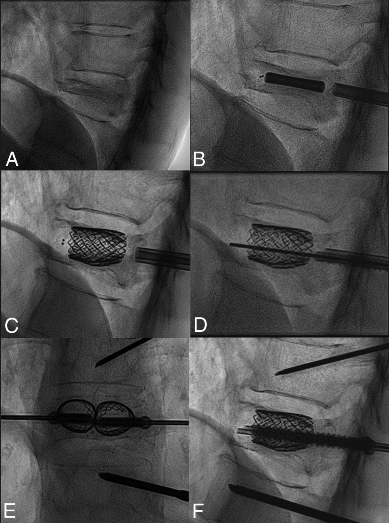
- FIG 4.
Standing plain film (A) and sagittal CT (B) show a T12 VP with pseudoarthrosis, gas cleft, and fracture mobility. Lateral intraprocedural fluoroscopic images before (C) and after (D) stent expansion with consequent fracture reduction. Postoperative standing plain film (E) demonstrates T12 height restoration and kyphosis correction, stable at 6 months’ follow-up (F). Axial CT (F) at the T12 level shows the stent-cement complex reconstructing the vertebral body and the transpedicular screws cemented inside the stents acting as “anchors” to the posterior elements.

Tables
Radiologic outcome: median measurements of anterior, middle, and posterior VBH, LKA, and VKA pre- and postoperatively (with IQR), for all fractures, mobile and nonmobile fracture groups
Preoperative (IQR) Postoperative (IQR) Median Gain Correction Loss at 6 Months (IQR) Ant VBHAll 9.5 mm (8.0–13.0) 17 mm (15.0–19.25) 7 mm, +74% (P < .001) Mobile 11.5 mm (9.0–15.25) 18 mm (16.5–19.5) 7 mm, +64% (P < .001) Nonmobile 11 mm (5.5–12.5) 19 mm (16.5–17.5) 8 mm, +73% (P = .03) Mid VBHAll 6 mm (5.0–7.75) 15.5 mm (13.0–17.25) 9 mm, +150% (P < .001) Mobile 6.5 mm (5.75–9.5) 16 mm (15.0–18.0) 9 mm, +138% (P < .001) Nonmobile 5 mm (4.5-5.5) 15.5 mm (13.5–16.75) 11 mm, +220% (P = .03) Post VBHAll 17.5 mm (16.0–19.0) 20 mm (18.0–22.0) 3 mm, +17% (P < .001) Mobile 18 mm (16.0–19.25) 20 mm (18.5–23.5) 3 mm, +17% (P < .001) Nonmobile 17 mm (15.0–18.0) 21 mm (20.0–22.0) 4.5 mm, +26% (P = .04) LKAAll 25° (12.0–29.0) 14° (6.0–22.0) 8° (P < .001) 1° (0.0–1.0) Mobile 25° (15.5–31.5) 14° (6.0–22.0) 8° (P < .001) 1° (0.0–1.7) Nonmobile 21.5° (11.25–27.75) 13° (4.5–15.0) 4.5° (P = .009) 1° (0.5–1.0) VKAAll 21° (12.0–27.0) 9° (5.5–12.0) 10° (P < .001) 0° (0.0–1.0) Mobile 23° (12.0–27.0) 9° (5.5–12.0) 11° (P < .001) 0° (0.0–1.0) Nonmobile 19.5° (13.25–22.5) 7° (3.75–12.0) 9.5° (P = .006) 0° (0.0–1.0)


