Advertisement

Main menu

  • Home
  • Content
    • Current Issue
    • Accepted Manuscripts
    • Article Preview
    • Past Issue Archive
    • Video Articles
    • AJNR Case Collection
    • Case of the Week Archive
    • Case of the Month Archive
    • Classic Case Archive
  • Special Collections
    • AJNR Awards
    • ASNR Foundation Special Collection
    • Most Impactful AJNR Articles
    • Photon-Counting CT
    • Spinal CSF Leak Articles (Jan 2020-June 2024)
  • Multimedia
    • AJNR Podcasts
    • AJNR SCANtastic
    • Trainee Corner
    • MRI Safety Corner
    • Imaging Protocols
  • For Authors
    • Submit a Manuscript
    • Submit a Video Article
    • Submit an eLetter to the Editor/Response
    • Manuscript Submission Guidelines
    • Statistical Tips
    • Fast Publishing of Accepted Manuscripts
    • Graphical Abstract Preparation
    • Imaging Protocol Submission
    • Author Policies
  • About Us
    • About AJNR
    • Editorial Board
    • Editorial Board Alumni
  • More
    • Become a Reviewer/Academy of Reviewers
    • Subscribers
    • Permissions
    • Alerts
    • Feedback
    • Advertisers
    • ASNR Home

User menu

  • Alerts
  • Log in

Search

  • Advanced search
American Journal of Neuroradiology

American Journal of Neuroradiology

ASHNR American Society of Functional Neuroradiology ASHNR American Society of Pediatric Neuroradiology ASSR
  • Alerts
  • Log in

Advanced Search

  • Home
  • Content
    • Current Issue
    • Accepted Manuscripts
    • Article Preview
    • Past Issue Archive
    • Video Articles
    • AJNR Case Collection
    • Case of the Week Archive
    • Case of the Month Archive
    • Classic Case Archive
  • Special Collections
    • AJNR Awards
    • ASNR Foundation Special Collection
    • Most Impactful AJNR Articles
    • Photon-Counting CT
    • Spinal CSF Leak Articles (Jan 2020-June 2024)
  • Multimedia
    • AJNR Podcasts
    • AJNR SCANtastic
    • Trainee Corner
    • MRI Safety Corner
    • Imaging Protocols
  • For Authors
    • Submit a Manuscript
    • Submit a Video Article
    • Submit an eLetter to the Editor/Response
    • Manuscript Submission Guidelines
    • Statistical Tips
    • Fast Publishing of Accepted Manuscripts
    • Graphical Abstract Preparation
    • Imaging Protocol Submission
    • Author Policies
  • About Us
    • About AJNR
    • Editorial Board
    • Editorial Board Alumni
  • More
    • Become a Reviewer/Academy of Reviewers
    • Subscribers
    • Permissions
    • Alerts
    • Feedback
    • Advertisers
    • ASNR Home
  • Follow AJNR on Twitter
  • Visit AJNR on Facebook
  • Follow AJNR on Instagram
  • Join AJNR on LinkedIn
  • RSS Feeds

AJNR is seeking candidates for the AJNR Podcast Editor. Read the position description.

Research ArticleInterventional
Open Access

Integrating 3D Rotational Angiography into Gamma Knife Planning

H. Hasegawa, S. Hanakita, M. Shin, M. Kawashima, T. Kin, W. Takahashi, M. Shojima, A.K. Nomoto, S. Aoki and N. Saito
American Journal of Neuroradiology October 2018, 39 (10) 1867-1870; DOI: https://doi.org/10.3174/ajnr.A5763
H. Hasegawa
aFrom the Departments of Neurosurgery (H.H., S.H., M. Shin, M.K., T.K., M. Shojima, N.S.)
  • Find this author on Google Scholar
  • Find this author on PubMed
  • Search for this author on this site
  • ORCID record for H. Hasegawa
S. Hanakita
aFrom the Departments of Neurosurgery (H.H., S.H., M. Shin, M.K., T.K., M. Shojima, N.S.)
  • Find this author on Google Scholar
  • Find this author on PubMed
  • Search for this author on this site
  • ORCID record for S. Hanakita
M. Shin
aFrom the Departments of Neurosurgery (H.H., S.H., M. Shin, M.K., T.K., M. Shojima, N.S.)
  • Find this author on Google Scholar
  • Find this author on PubMed
  • Search for this author on this site
  • ORCID record for M. Shin
M. Kawashima
aFrom the Departments of Neurosurgery (H.H., S.H., M. Shin, M.K., T.K., M. Shojima, N.S.)
  • Find this author on Google Scholar
  • Find this author on PubMed
  • Search for this author on this site
  • ORCID record for M. Kawashima
T. Kin
aFrom the Departments of Neurosurgery (H.H., S.H., M. Shin, M.K., T.K., M. Shojima, N.S.)
  • Find this author on Google Scholar
  • Find this author on PubMed
  • Search for this author on this site
  • ORCID record for T. Kin
W. Takahashi
bRadiology (W.T., A.K.N., S.A.), University of Tokyo Hospital, Tokyo, Japan.
  • Find this author on Google Scholar
  • Find this author on PubMed
  • Search for this author on this site
  • ORCID record for W. Takahashi
M. Shojima
aFrom the Departments of Neurosurgery (H.H., S.H., M. Shin, M.K., T.K., M. Shojima, N.S.)
  • Find this author on Google Scholar
  • Find this author on PubMed
  • Search for this author on this site
  • ORCID record for M. Shojima
A.K. Nomoto
bRadiology (W.T., A.K.N., S.A.), University of Tokyo Hospital, Tokyo, Japan.
  • Find this author on Google Scholar
  • Find this author on PubMed
  • Search for this author on this site
  • ORCID record for A.K. Nomoto
S. Aoki
bRadiology (W.T., A.K.N., S.A.), University of Tokyo Hospital, Tokyo, Japan.
  • Find this author on Google Scholar
  • Find this author on PubMed
  • Search for this author on this site
  • ORCID record for S. Aoki
N. Saito
aFrom the Departments of Neurosurgery (H.H., S.H., M. Shin, M.K., T.K., M. Shojima, N.S.)
  • Find this author on Google Scholar
  • Find this author on PubMed
  • Search for this author on this site
  • ORCID record for N. Saito
  • Article
  • Figures & Data
  • Supplemental
  • Info & Metrics
  • Responses
  • References
  • PDF
Loading

Abstract

SUMMARY: 3D rotational angiography provides remarkable spatial resolution for cerebrovascular disorders; however, it cannot be integrated directly into gamma knife planning due to the discrepancy of DICOM “tag” information, and most physicians still cannot benefit from 3D rotational angiography. Here, we describe a simple and easy technique to enable the integration of 3D rotational angiography.

ABBREVIATIONS:

GKRS
gamma knife stereotactic radiosurgery
3DRA
3D rotational angiography

Gamma knife stereotactic radiosurgery (GKRS) is an image-guided radiation therapy characterized by its high geometric accuracy; thus, no treatment margin is usually required when circumscribing the target. This feature, in conjunction with its sharp dose fall-off, enables high-dose irradiation in a single session; however, successful radiosurgery is highly dependent on the quality of radiographic images used.

GKRS has been accepted as one of the standard therapeutic modalities for small-to-medium arteriovenous malformations.1⇓⇓⇓⇓⇓⇓⇓–9 Currently, biplanar DSA and CT angiography or MR imaging or both are commonly used in most institutions.10⇓⇓⇓⇓–15 Recently, advances in modern endovascular suites and newer generation flat panel detectors with C-arm systems have enabled acquisition of 3D rotational angiography (3DRA), providing remarkable spatial resolution for cerebrovascular disorders. Indeed, 3DRA is beginning to be used in the treatment planning of other modalities of stereotactic radiation therapy, including CyberKnife (Accuray, Sunnyvale, California), XKnife (Integra LifeSciences, Plainsboro, New Jersey), and Trilogy (Varian Medical Systems, Palo Alto, California), contributing to improved accuracy of the treatment planning.16⇓–18 One remaining issue is that 3DRA cannot be integrated directly into GKRS planning because the planning software (Leksell GammaPlan; Elekta Instruments, Stockholm, Sweden) does not accept 3DRA. Moreover, very few studies have described the effectiveness of 3DRA on GKRS planning, though they do not state a detailed integration method.19⇓–21 Thus, most physicians still cannot benefit from 3DRA. Here, we show a very simple method to integrate 3DRA into GKRS planning.

Techniques

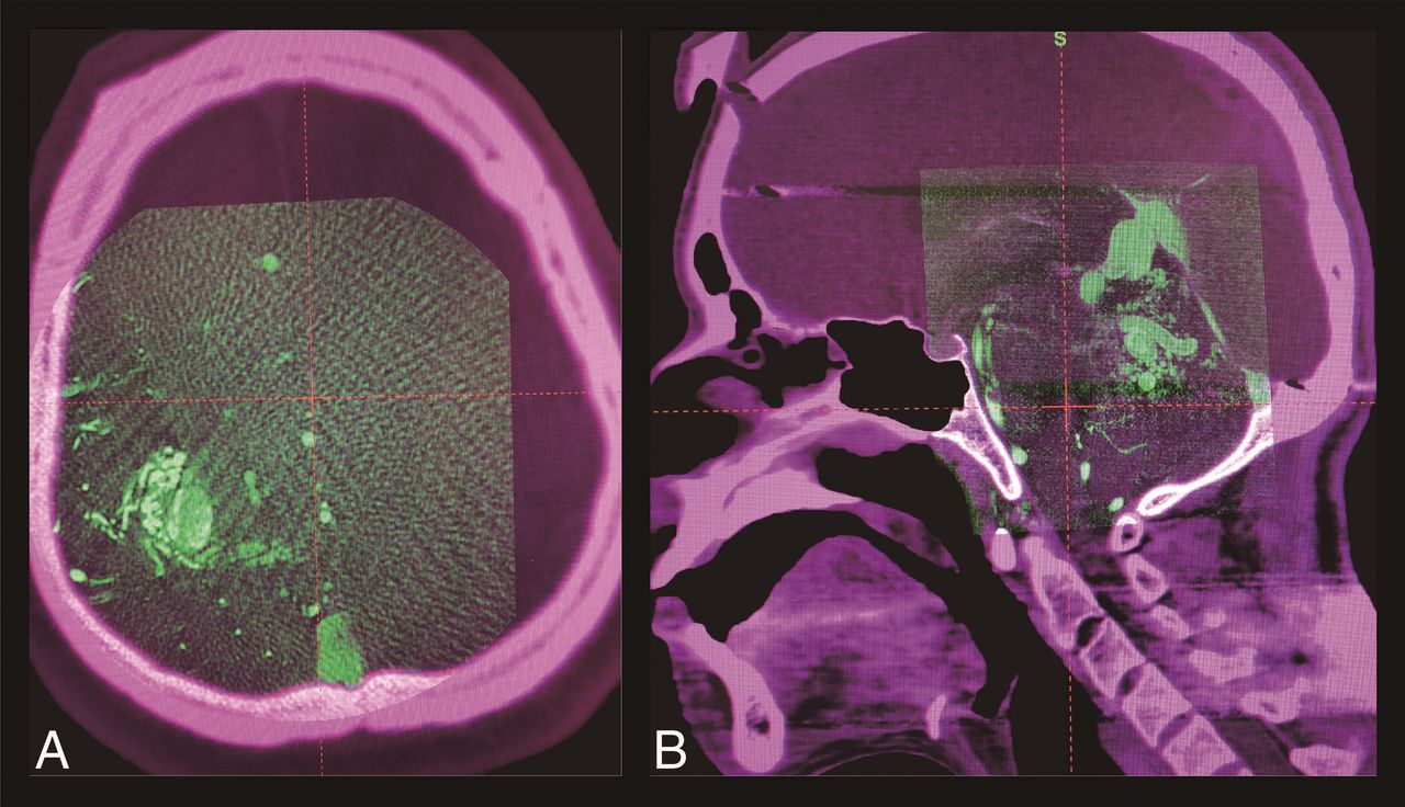
Before the day of treatment, we usually perform MR imaging (mainly time-of-flight MR angiography, supplemented by T2 and gadolinium-enhanced T1 images) for preplanning. As in the usual preparations for GKRS, the Leksell frame (Elekta Instruments) is set on the patient's head with the patient under sedation with local anesthesia. Then, the patient is transferred to the angiographic suite (Allura Xper FD20/10; Philips Healthcare, Best, Netherlands). Along with conventional DSA, 3DRA is acquired using the programmed acquisition protocol (3DRA mode). The amount of contrast medium used and preinjection delay are individually determined by neuroendovascular surgeons; briefly, 1–3 mL/s of contrast medium is continuously injected with a 1.5- to 2.0-second preinjection delay during the rotation of the C-arm. Because 3DRA could be easily coregistered to stereotactic CT if 3DRA contained enough bony tissue, a large-sized detector is generally preferred for precise image coregistration (Fig 1A). On the contrary, the smallest 8-inch detector could be used when the nidus is located near the skull base (basal ganglia, posterior fossa, and so forth) because acquired images spontaneously contain a large portion of bony tissues (Fig 1B). The obtained volume dataset is automatically transferred to the preinstalled workstation (XtraVision; Philips Healthcare), with which further reconstruction is performed with a 2563- or 5123-resolution voxel matrix in a planned cube-shaped FOV with preset side lengths of 34.96, 52.18, 69.92, or 104.36 mm. We mostly use a 69.92-mm side-length cube with a 2563-resolution voxel matrix to maintain the balance between spatial resolution and contrast resolution; thus, spatial resolution is roughly calculated as 0.27 mm. Then, the DICOM “tag” technique is corrected from XA into CT in our software so that the GammaPlan can recognize the 3DRA properly as a CT-like image. Once the 3DRA is installed in the GammaPlan, image coregistration to stereotactic CT will be automated using the preinstalled coregistration function. A step-by-step instruction manual to integrate 3DRA is shown in On-line Figure. Detailed case illustrations are shown in Fig 2.

Fig 1.
  • Download figure
  • Open in new tab
  • Download powerpoint
Fig 1.

3D rotational angiography images obtained with a large (A) and an 8-inch detector (B) are coregistered to each stereotactic CT image on treatment-planning software (GammaPlan).

Fig 2.
  • Download figure
  • Open in new tab
  • Download powerpoint
Fig 2.

A 34-year-old woman with an unruptured, small, left medial frontal arteriovenous malformation, once treated with gamma knife (A). The small remnant nidus persisted at 3.5 years from the initial radiosurgery (B, a red arrowhead shows the remnant nidus). The actual treatment planning of the secondary treatment (C) shows that 3D rotational angiography (left column) successfully depicts the faint remnant, while both time-of-flight (middle column) and gadolinium-enhanced T1 images (right column) fail to depict it. The nidus was finally obliterated (D) at 1.5 years from the secondary treatment. Yellow lines show the prescription isodose lines.

Discussion

With the above-described simple contrivance, 3DRA can readily be used for GKRS planning. The strength of this technique is its accessibility. GammaPlan cannot directly accept 3DRA because of the discrepancy of DICOM tag information; accordingly, we must address this issue. No changes are required in the geometric or patient information; thus, the image quality itself is intact. Because 3DRA is a CT-like image, being characteristic of the finest spatial resolution for high-contrast objects with poor contrast resolution and thus having many features common to CT,22,23 stereotactic CT would be the best reference image for the coregistration.18 By means of high-definition images, physicians can not only reduce unwanted waste radiation to the surrounding brain tissues but also enable a safe prescription of high radiosurgical doses, enough to obliterate the nidus, which might theoretically lead to improvement in the obliteration rate as well as a decrease in radiation-induced adverse events. However, this article is only a technical report, and the actual clinical outcomes should be further examined.

Although 3DRA mode is preferred in our institution, contrast-enhanced conebeam CT with a small targeted FOV (high-resolution XperCT mode; Philips Healthcare) is also available in our angiographic suite. This acquisition mode provides superior spatial resolution with a long acquisition time (20 seconds), using a slow C-arm rotation and an 8-inch detector, which might be better for very small arteriovenous malformations. However, use of the high-resolution XperCT mode might raise a concern about coregistration. Notably, images containing a large portion of the cranium as well as skull base bone are important for precise coregistration; the use of a smaller detector contributes to further increased spatial resolution but also loses images of the surrounding cranium and skull base, leading to difficulty in coregistration. We usually perform stereotactic CT after finishing angiography so that the contrast medium remaining in the blood vessels can provide additional information of vascular anatomies, which may enhance the quality of image coregistration. Further research is desirable to examine the coregistration accuracy to ensure the quality of the prescription of the therapeutic radiation dose.

Moreover, when a nidus receives blood flow from >2 vessels, the precise nidus contour is shown as the summation of parts of the nidus obtained by cannulation in each vessel. Thus, it is quite important to perform 3- or 4-vessel angiography and judge the involvement to avoid underestimation of the whole nidus angioarchitecture.

Although 3DRA provides superior resolution for angioarchitectures of vascular lesions, we recommend creating radiosurgical plans by meticulously comparing all the available imaging modalities. Particularly, MR imaging exhibits excellent contrast resolution and provides a better understanding of the surrounding functional brain anatomies, and DSA enables surgeons to instinctively recognize a spatial expanse of the nidus. Notably, surgeons should manipulate the DICOM header at their own risk because carelessly manipulating DICOM could spoil the quality of the images. Meticulous care must be taken not to change important information in the DICOM header other than technique because it could alter spatial relationships. Given the above reasons, 3DRA should not be used as a main technique but as reference information during radiosurgical planning.

Conclusions

We describe a simple, easy-to-access technique to enable integration of 3DRA into GKRS planning, which could provide the highest resolution for angioarchitectures of vascular lesions. The present method remains preliminary; thus, the created treatment plans should be validated in comparison with the conventional planning method. Further research is desirable to assess the effect of this technique on the actual radiosurgical outcomes.

Footnotes

  • Disclosures: Hirotaka Hasegawa—RELATED: Grant: JSPS KAKENHI, Comments: grant No. JP17K16628.

  • This study was supported by Japan Society for the Promotion of Science (JSPS) KAKENHI grant (No. JP17K16628).

Indicates open access to non-subscribers at www.ajnr.org

References

  1. 1.↵
    1. Cohen-Gadol AA,
    2. Pollock BE
    . Radiosurgery for arteriovenous malformations in children. J Neurosurg 2006;104:388–91 pmid:16776373
    PubMed
  2. 2.↵
    1. Lunsford LD,
    2. Kondziolka D,
    3. Flickinger JC, et al
    . Stereotactic radiosurgery for arteriovenous malformations of the brain. J Neurosurg 1991;75:512–24 doi:10.3171/jns.1991.75.4.0512 pmid:1885968
    CrossRefPubMed
  3. 3.↵
    1. Karlsson B,
    2. Lindquist C,
    3. Steiner L
    . Prediction of obliteration after gamma knife surgery for cerebral arteriovenous malformations. Neurosurgery 1997;40:425–30; discussion 430–31 pmid:9055280
    CrossRefPubMed
  4. 4.↵
    1. Schwartz M,
    2. Sixel K,
    3. Young C, et al
    . Prediction of obliteration of arteriovenous malformations after radiosurgery: the obliteration prediction index. Can J Neurol Sci 1997;24:106–09 doi:10.1017/S0317167100021417 pmid:9164685
    CrossRefPubMed
  5. 5.↵
    1. Flickinger JC,
    2. Kondziolka D,
    3. Maitz AH, et al
    . An analysis of the dose-response for arteriovenous malformation radiosurgery and other factors affecting obliteration. Radiother Oncol 2002;63:347–54 doi:10.1016/S0167-8140(02)00103-2 pmid:12142099
    CrossRefPubMed
  6. 6.↵
    1. Fleetwood IG,
    2. Steinberg GK
    . Arteriovenous malformations. Lancet 2002;359:863–73 doi:10.1016/S0140-6736(02)07946-1 pmid:11897302
    CrossRefPubMed
  7. 7.↵
    1. Shin M,
    2. Kawamoto S,
    3. Kurita H, et al
    . Retrospective analysis of a 10-year experience of stereotactic radio surgery for arteriovenous malformations in children and adolescents. J Neurosurg 2002;97:779–84 doi:10.3171/jns.2002.97.4.0779 pmid:12405363
    CrossRefPubMed
  8. 8.↵
    1. Starke RM,
    2. Kano H,
    3. Ding D, et al
    . Stereotactic radiosurgery for cerebral arteriovenous malformations: evaluation of long-term outcomes in a multicenter cohort. J Neurosurg 2017;126:36–44 doi:10.3171/2015.9.JNS151311 pmid:26943847
    CrossRefPubMed
  9. 9.↵
    1. Starke RM,
    2. Ding D,
    3. Kano H, et al
    . International multicenter cohort study of pediatric brain arteriovenous malformations, part 2: outcomes after stereotactic radiosurgery. J Neurosurg Pediatr 2017;19:136–48 doi:10.3171/2016.9.PEDS16284 pmid:27911249
    CrossRefPubMed
  10. 10.↵
    1. Blatt DR,
    2. Friedman WA,
    3. Bova FJ
    . Modifications based on computed tomographic imaging in planning the radiosurgical treatment of arteriovenous malformations. Neurosurgery 1993;33:588–95; discussion 595–96 pmid:8232797
    PubMed
  11. 11.↵
    1. Ding D,
    2. Xu Z,
    3. Shih HH, et al
    . Stereotactic radiosurgery for partially resected cerebral arteriovenous malformations. World Neurosurg 2016;85:263–72 doi:10.1016/j.wneu.2015.10.001 pmid:26459698
    CrossRefPubMed
  12. 12.↵
    1. Hanakita S,
    2. Koga T,
    3. Shin M, et al
    . The long-term outcomes of radiosurgery for arteriovenous malformations in pediatric and adolescent populations. J Neurosurg Pediatr 2015;16:222–31 doi:10.3171/2015.1.PEDS14407 pmid:25955806
    CrossRefPubMed
  13. 13.↵
    1. Kano H,
    2. Lunsford LD,
    3. Flickinger JC, et al
    . Stereotactic radiosurgery for arteriovenous malformations, part 1: management of Spetzler-Martin Grade I and II arteriovenous malformations. J Neurosurg 2012;116:11–20 doi:10.3171/2011.9.JNS101740 pmid:22077452
    CrossRefPubMed
  14. 14.↵
    1. Kondziolka D,
    2. Lunsford LD,
    3. Kanal E, et al
    . Stereotactic magnetic resonance angiography for targeting in arteriovenous malformation radiosurgery. Neurosurgery 1994;35:585–90; discussion 590–91 doi:10.1227/00006123-199410000-00002 pmid:7808600
    CrossRefPubMed
  15. 15.↵
    1. Lunsford LD,
    2. Niranjan A,
    3. Kano H, et al
    . The technical evolution of gamma knife radiosurgery for arteriovenous malformations. Prog Neurol Surg 2013;27:22–34 doi:10.1159/000341625 pmid:23258506
    CrossRefPubMed
  16. 16.↵
    1. van der Bom IM,
    2. Gounis MJ,
    3. Ding L, et al
    . Target delineation for radiosurgery of a small brain arteriovenous malformation using high-resolution contrast-enhanced cone beam CT. BMJ Case Rep 2013;2013 doi:10.1136/bcr-2013-010763 pmid:23946527
    CrossRefPubMed
  17. 17.↵
    1. Colombo F,
    2. Cavedon C,
    3. Francescon P, et al
    . Three-dimensional angiography for radiosurgical treatment planning for arteriovenous malformations. J Neurosurg 2003;98:536–43 doi:10.3171/jns.2003.98.3.0536 pmid:12650425
    CrossRefPubMed
  18. 18.↵
    1. Hristov D,
    2. Liu L,
    3. Adler JR, et al
    . Technique for targeting arteriovenous malformations using frameless image-guided robotic radiosurgery. Int J Radiat Oncol Biol Phys 2011;79:1232–40 doi:10.1016/j.ijrobp.2010.05.015 pmid:20801584
    CrossRefPubMed
  19. 19.↵
    1. Safain MG,
    2. Rahal JP,
    3. Raval A, et al
    . Use of cone-beam computed tomography angiography in planning for gamma knife radiosurgery for arteriovenous malformations: a case series and early report. Neurosurgery 2014;74:682–95; discussion 695–96 doi:10.1227/NEU.0000000000000331 pmid:24584136
    CrossRefPubMed
  20. 20.↵
    1. Kang J,
    2. Huang J,
    3. Gailloud P, et al
    . Planning evaluation of C-arm cone beam CT angiography for target delineation in stereotactic radiation surgery of brain arteriovenous malformations. Int J Radiat Oncol Biol Phys 2014;90:430–37 doi:10.1016/j.ijrobp.2014.05.035 pmid:25015197
    CrossRefPubMed
  21. 21.↵
    1. Chen KK,
    2. Guo WY,
    3. Yang HC, et al
    . Application of time-resolved 3D digital subtraction angiography to plan cerebral arteriovenous malformation radiosurgery. AJNR Am J Neuroradiol 2017;38:740–46 doi:10.3174/ajnr.A5074 pmid:28126751
    Abstract/FREE Full Text
  22. 22.↵
    1. Miracle AC,
    2. Mukherji SK
    . Conebeam CT of the head and neck, part 1: physical principles. AJNR Am J Neuroradiol 2009;30:1088–95 doi:10.3174/ajnr.A1653 pmid:19439484
    Abstract/FREE Full Text
  23. 23.↵
    1. Kanayama S,
    2. Hara T,
    3. Hamada Y, et al
    . Potential of 80-kV high-resolution cone-beam CT imaging combined with an optimized protocol for neurological surgery. Neuroradiology 2015;57:155–62 doi:10.1007/s00234-014-1447-7 pmid:25370477
    CrossRefPubMed
  • Received April 4, 2018.
  • Accepted after revision June 12, 2018.
  • © 2018 by American Journal of Neuroradiology
View Abstract
PreviousNext
Back to top

In this issue

American Journal of Neuroradiology: 39 (10)
American Journal of Neuroradiology
Vol. 39, Issue 10
1 Oct 2018
  • Table of Contents
  • Index by author
  • Complete Issue (PDF)
Advertisement
Print
Download PDF
Email Article

Thank you for your interest in spreading the word on American Journal of Neuroradiology.

NOTE: We only request your email address so that the person you are recommending the page to knows that you wanted them to see it, and that it is not junk mail. We do not capture any email address.

Enter multiple addresses on separate lines or separate them with commas.
Integrating 3D Rotational Angiography into Gamma Knife Planning
(Your Name) has sent you a message from American Journal of Neuroradiology
(Your Name) thought you would like to see the American Journal of Neuroradiology web site.
CAPTCHA
This question is for testing whether or not you are a human visitor and to prevent automated spam submissions.
Cite this article
H. Hasegawa, S. Hanakita, M. Shin, M. Kawashima, T. Kin, W. Takahashi, M. Shojima, A.K. Nomoto, S. Aoki, N. Saito
Integrating 3D Rotational Angiography into Gamma Knife Planning
American Journal of Neuroradiology Oct 2018, 39 (10) 1867-1870; DOI: 10.3174/ajnr.A5763

Citation Manager Formats

  • BibTeX
  • Bookends
  • EasyBib
  • EndNote (tagged)
  • EndNote 8 (xml)
  • Medlars
  • Mendeley
  • Papers
  • RefWorks Tagged
  • Ref Manager
  • RIS
  • Zotero
0 Responses
Respond to this article
Share
Bookmark this article
Integrating 3D Rotational Angiography into Gamma Knife Planning
H. Hasegawa, S. Hanakita, M. Shin, M. Kawashima, T. Kin, W. Takahashi, M. Shojima, A.K. Nomoto, S. Aoki, N. Saito
American Journal of Neuroradiology Oct 2018, 39 (10) 1867-1870; DOI: 10.3174/ajnr.A5763
del.icio.us logo Twitter logo Facebook logo Mendeley logo
  • Tweet Widget
  • Facebook Like
  • Google Plus One
Purchase

Jump to section

  • Article
    • Abstract
    • ABBREVIATIONS:
    • Discussion
    • Conclusions
    • Footnotes
    • References
  • Figures & Data
  • Supplemental
  • Info & Metrics
  • Responses
  • References
  • PDF

Related Articles

  • PubMed
  • Google Scholar

Cited By...

  • No citing articles found.
  • Crossref (6)
  • Google Scholar

This article has been cited by the following articles in journals that are participating in Crossref Cited-by Linking.

  • Rotational Angiography-Based Gamma Knife Radiosurgery for Brain Arteriovenous Malformations: Preliminary Therapeutic Outcomes of the Novel Method
    Yuki Shinya, Hirotaka Hasegawa, Masahiro Shin, Mariko Kawashima, Takehiro Sugiyama, Osamu Ishikawa, Satoshi Koizumi, Yuichi Suzuki, Hirofumi Nakatomi, Nobuhito Saito
    Neurosurgery 2021 89 1
  • Prognosis of Rotational Angiography-Based Stereotactic Radiosurgery for Dural Arteriovenous Fistulas: A Retrospective Analysis
    Yuki Shinya, Hirotaka Hasegawa, Mariko Kawashima, Satoshi Koizumi, Atsuto Katano, Motoyuki Umekawa, Nobuhito Saito
    Neurosurgery 2023 92 1
  • Recent Advances and Controversies in Gamma Knife Neurosurgery
    Jeremy C. Ganz
    2022 268
  • Aplicaciones de la tomografía computarizada de haz cónico en procedimientos mínimamente invasivos guiados por imagen
    E. Serrano, J. Valcárcel José, A. Páez-Carpio, M. Matute-González, M.F. Werner, A. López-Rueda
    Radiología 2025 67 1
  • Cone Beam computed tomography (CBCT) applications in image-guided minimally invasive procedures
    E. Serrano, J. Valcárcel José, A. Páez-Carpio, M. Matute-González, M.F. Werner, A. López-Rueda
    Radiología (English Edition) 2025 67 1
  • Evaluation of micro-remnant niduses of arteriovenous malformations post-gamma knife radiosurgery by 3D-rotational angiography
    Ryuichi Noda, Atsuya Akabane, Mariko Kawashima, Masafumi Segawa, Sho Tsunoda, Hiroyuki Wada, Makoto Watanabe, Haruyasu Yamada, Tomohiro Inoue
    Acta Neurochirurgica 2024 166 1

More in this TOC Section

  • SAVE vs. Solumbra Techniques for Thrombectomy
  • Contrast-Induced Encephalopathy after NeuroIR
  • CT Perfusion&Reperfusion in Acute Ischemic Stroke
Show more Interventional

Similar Articles

Advertisement

Indexed Content

  • Current Issue
  • Accepted Manuscripts
  • Article Preview
  • Past Issues
  • Editorials
  • Editor's Choice
  • Fellows' Journal Club
  • Letters to the Editor
  • Video Articles

Cases

  • Case Collection
  • Archive - Case of the Week
  • Archive - Case of the Month
  • Archive - Classic Case

Special Collections

  • AJNR Awards
  • ASNR Foundation Special Collection
  • Most Impactful AJNR Articles
  • Photon-Counting CT
  • Spinal CSF Leak Articles (Jan 2020-June 2024)

More from AJNR

  • Trainee Corner
  • Imaging Protocols
  • MRI Safety Corner

Multimedia

  • AJNR Podcasts
  • AJNR Scantastics

Resources

  • Turnaround Time
  • Submit a Manuscript
  • Submit a Video Article
  • Submit an eLetter to the Editor/Response
  • Manuscript Submission Guidelines
  • Statistical Tips
  • Fast Publishing of Accepted Manuscripts
  • Graphical Abstract Preparation
  • Imaging Protocol Submission
  • Evidence-Based Medicine Level Guide
  • Publishing Checklists
  • Author Policies
  • Become a Reviewer/Academy of Reviewers
  • News and Updates

About Us

  • About AJNR
  • Editorial Board
  • Editorial Board Alumni
  • Alerts
  • Permissions
  • Not an AJNR Subscriber? Join Now
  • Advertise with Us
  • Librarian Resources
  • Feedback
  • Terms and Conditions
  • AJNR Editorial Board Alumni

American Society of Neuroradiology

  • Not an ASNR Member? Join Now

© 2025 by the American Society of Neuroradiology All rights, including for text and data mining, AI training, and similar technologies, are reserved.
Print ISSN: 0195-6108 Online ISSN: 1936-959X

Powered by HighWire