Advertisement

Main menu

  • Home
  • Content
    • Current Issue
    • Accepted Manuscripts
    • Article Preview
    • Past Issue Archive
    • Video Articles
    • AJNR Case Collection
    • Case of the Week Archive
    • Case of the Month Archive
    • Classic Case Archive
  • Special Collections
    • AJNR Awards
    • ASNR Foundation Special Collection
    • Most Impactful AJNR Articles
    • Photon-Counting CT
    • Spinal CSF Leak Articles (Jan 2020-June 2024)
  • Multimedia
    • AJNR Podcasts
    • AJNR SCANtastic
    • Trainee Corner
    • MRI Safety Corner
    • Imaging Protocols
  • For Authors
    • Submit a Manuscript
    • Submit a Video Article
    • Submit an eLetter to the Editor/Response
    • Manuscript Submission Guidelines
    • Statistical Tips
    • Fast Publishing of Accepted Manuscripts
    • Graphical Abstract Preparation
    • Imaging Protocol Submission
    • Author Policies
  • About Us
    • About AJNR
    • Editorial Board
    • Editorial Board Alumni
  • More
    • Become a Reviewer/Academy of Reviewers
    • Subscribers
    • Permissions
    • Alerts
    • Feedback
    • Advertisers
    • ASNR Home

User menu

  • Alerts
  • Log in

Search

  • Advanced search
American Journal of Neuroradiology

American Journal of Neuroradiology

ASHNR American Society of Functional Neuroradiology ASHNR American Society of Pediatric Neuroradiology ASSR
  • Alerts
  • Log in

Advanced Search

  • Home
  • Content
    • Current Issue
    • Accepted Manuscripts
    • Article Preview
    • Past Issue Archive
    • Video Articles
    • AJNR Case Collection
    • Case of the Week Archive
    • Case of the Month Archive
    • Classic Case Archive
  • Special Collections
    • AJNR Awards
    • ASNR Foundation Special Collection
    • Most Impactful AJNR Articles
    • Photon-Counting CT
    • Spinal CSF Leak Articles (Jan 2020-June 2024)
  • Multimedia
    • AJNR Podcasts
    • AJNR SCANtastic
    • Trainee Corner
    • MRI Safety Corner
    • Imaging Protocols
  • For Authors
    • Submit a Manuscript
    • Submit a Video Article
    • Submit an eLetter to the Editor/Response
    • Manuscript Submission Guidelines
    • Statistical Tips
    • Fast Publishing of Accepted Manuscripts
    • Graphical Abstract Preparation
    • Imaging Protocol Submission
    • Author Policies
  • About Us
    • About AJNR
    • Editorial Board
    • Editorial Board Alumni
  • More
    • Become a Reviewer/Academy of Reviewers
    • Subscribers
    • Permissions
    • Alerts
    • Feedback
    • Advertisers
    • ASNR Home
  • Follow AJNR on Twitter
  • Visit AJNR on Facebook
  • Follow AJNR on Instagram
  • Join AJNR on LinkedIn
  • RSS Feeds

AJNR is seeking candidates for the AJNR Podcast Editor. Read the position description.

Review ArticleREVIEW ARTICLE
Open Access

Resting-State Functional MRI: Everything That Nonexperts Have Always Wanted to Know

H. Lv, Z. Wang, E. Tong, L.M. Williams, G. Zaharchuk, M. Zeineh, A.N. Goldstein-Piekarski, T.M. Ball, C. Liao and M. Wintermark
American Journal of Neuroradiology January 2018, DOI: https://doi.org/10.3174/ajnr.A5527
H. Lv
aFrom the Department of Radiology (H.L., Z.W.), Beijing Friendship Hospital, Capital Medical University, Beijing, China
bDepartment of Radiology (H.L., G.Z., M.Z., M.W.), Neuroradiology Division
  • Find this author on Google Scholar
  • Find this author on PubMed
  • Search for this author on this site
  • ORCID record for H. Lv
Z. Wang
aFrom the Department of Radiology (H.L., Z.W.), Beijing Friendship Hospital, Capital Medical University, Beijing, China
  • Find this author on Google Scholar
  • Find this author on PubMed
  • Search for this author on this site
  • ORCID record for Z. Wang
E. Tong
dDepartment of Radiology (E.T.), Neuroradiology Section, University of California, San Francisco, San Francisco, California
  • Find this author on Google Scholar
  • Find this author on PubMed
  • Search for this author on this site
  • ORCID record for E. Tong
L.M. Williams
cDepartment of Psychiatry and Behavioral Sciences (L.M.W., A.N.G.-P., T.M.B.), Stanford University, Stanford, California
  • Find this author on Google Scholar
  • Find this author on PubMed
  • Search for this author on this site
  • ORCID record for L.M. Williams
G. Zaharchuk
bDepartment of Radiology (H.L., G.Z., M.Z., M.W.), Neuroradiology Division
  • Find this author on Google Scholar
  • Find this author on PubMed
  • Search for this author on this site
  • ORCID record for G. Zaharchuk
M. Zeineh
bDepartment of Radiology (H.L., G.Z., M.Z., M.W.), Neuroradiology Division
  • Find this author on Google Scholar
  • Find this author on PubMed
  • Search for this author on this site
  • ORCID record for M. Zeineh
A.N. Goldstein-Piekarski
cDepartment of Psychiatry and Behavioral Sciences (L.M.W., A.N.G.-P., T.M.B.), Stanford University, Stanford, California
  • Find this author on Google Scholar
  • Find this author on PubMed
  • Search for this author on this site
  • ORCID record for A.N. Goldstein-Piekarski
T.M. Ball
cDepartment of Psychiatry and Behavioral Sciences (L.M.W., A.N.G.-P., T.M.B.), Stanford University, Stanford, California
  • Find this author on Google Scholar
  • Find this author on PubMed
  • Search for this author on this site
  • ORCID record for T.M. Ball
C. Liao
eDepartment of Radiology (C.L.), Yunnan Tumor Hospital (The Third Affiliated Hospital of Kunming Medical University), Kunming, Yunnan Province, China.
  • Find this author on Google Scholar
  • Find this author on PubMed
  • Search for this author on this site
  • ORCID record for C. Liao
M. Wintermark
bDepartment of Radiology (H.L., G.Z., M.Z., M.W.), Neuroradiology Division
  • Find this author on Google Scholar
  • Find this author on PubMed
  • Search for this author on this site
  • ORCID record for M. Wintermark
  • Article
  • Figures & Data
  • Info & Metrics
  • Responses
  • References
  • PDF
Loading

Abstract

SUMMARY: Resting-state fMRI was first described by Biswal et al in 1995 and has since then been widely used in both healthy subjects and patients with various neurologic, neurosurgical, and psychiatric disorders. As opposed to paradigm- or task-based functional MR imaging, resting-state fMRI does not require subjects to perform any specific task. The low-frequency oscillations of the resting-state fMRI signal have been shown to relate to the spontaneous neural activity. There are many ways to analyze resting-state fMRI data. In this review article, we will briefly describe a few of these and highlight the advantages and limitations of each. This description is to facilitate the adoption and use of resting-state fMRI in the clinical setting, helping neuroradiologists become familiar with these techniques and applying them for the care of patients with neurologic and psychiatric diseases.

ABBREVIATIONS:

ALFF
Amplitude of Low Frequency Fluctuations
BOLD
blood oxygen level–dependent
FCD
functional connectivity density
ICA
independent component analysis
ReHo
regional homogeneity
rs-fMRI
resting-state fMRI

The brain controls all the complex functions in the human body. Structurally, the brain is organized grossly into different regions specialized for processing and relaying neural signals; functionally, the brain is subspecialized for perceptual and cognitive processes. Working in concert, these subspecialized areas orchestrate complex bodily functions and allow human behavior.

Neurons do not contain any internal reserves of energy, either in the form of glucose or oxygen. When activated, they are provided with more energy by the adjacent capillaries through a process called the hemodynamic response, which supplies them with increased regional cerebral blood flow and an increase in oxygen supply, usually even greater than their needs.1,2 This process results in a change in terms of the relative levels of oxyhemoglobin and deoxyhemoglobin that can be detected by MR imaging on the basis of their differential magnetic susceptibilities. This imaging approach is called blood oxygen level–dependent (BOLD) contrast imaging. Conventionally, BOLD signal change has been known to be modulated by the arterial partial pressure of CO2 level. More recent research suggests that BOLD signal is determined by both the arterial partial pressure of O2 and CO2, rather than CO2 alone.3 The change in the BOLD signal is the cornerstone of functional MR imaging,4,5 which is traditionally used to construct maps indicating subspecialized brain regions that are activated by certain tasks or reacting to a stimulus at a low frequency (0.01–0.1Hz). The frequencies of neural activity fluctuations measured by fMRI (which are low-frequency and indirectly measured using BOLD signal) and of neural firing measured in neurophysiologic studies (which are high-frequency and are directly measured) are different.6,7

The biologic significance of the neural activity fluctuations was first described by Biswal et al in 1995.8 When the subjects were asked to perform bilateral finger tapping in that experiment, researchers identified a highly correlated BOLD time course between the left somatosensory cortex and the homologous areas in the contralateral hemisphere.8 Since then, fMRI has been widely used in both healthy subjects and patients with neurologic and psychiatric disorders to analyze synchronous, spontaneous fluctuations of various resting-state networks.9 Because of the neurovascular coupling, the BOLD signal, while vascular in nature, is strongly related to neuronal activity.10 However, the delay of the hemodynamic response following neural activation is responsible for the relatively poor temporal resolution of fMRI,11 and the BOLD signal can be altered in brain regions where the blood circulation is altered.12⇓–14 For example, pathologic conditions such as traumatic brain injury15⇓–17 or anoxic brain injury18⇓⇓–21 might affect neurovascular coupling and therefore make fMRI suboptimal to assess neural activity in these pathologic conditions. Therefore, these factors should be taken into consideration when designing calibrated BOLD experiments and interpreting functional connectivity data, especially in patients with vascular pathologies.3

As opposed to paradigm- or task-based functional MR imaging, resting-state fMRI (rs-fMRI) is acquired in the absence of a stimulus or a task, in other words at rest. The principle of rs-fMRI is also based on the BOLD signal fluctuation, which is the same as for active-task fMRI. rs-fMRI focuses on spontaneous BOLD signal alterations. Data can be acquired with a dedicated scan, in which individuals are instructed to simply rest, or by inferring resting-state data from periods of rest embedded within a series of tasks.22 The lack of a task makes rs-fMRI particularly attractive for patients who may have difficulty with task instructions, such as those with neurologic, neurosurgical, and psychiatric conditions, as well as for pediatric patients. Thus, the application of rs-fMRI in the research and clinical setting has been growing for the past 2 decades.9,23

There are multiple ways to analyze rs-fMRI data, and each approach has implications in terms of what information can be extracted from the data. This article attempts to provide a broad overview of major analysis methods that are used in rs-fMRI. We will systematically describe these approaches, their advantages, and limitations. This work will help nonexperts become familiar with rs-fMRI techniques and how to apply them to the benefit of patients with neurologic, neurosurgical, and psychiatric disorders.24,25

Analytic Methods

rs-fMRI analysis is challenging due to the massive amount of data and the need for sophisticated analysis. Before one applies any of the analytic methods, realignment and removal of confounding artifacts (eg, head motion, CSF signal) are important preprocessing steps. Other main preprocessing steps are required on rs-fMRI data, including removing the first 10∼20 time points, slice timing, data normalization, and band-pass filtering. Other possible procedures, including smoothing, may be performed in different sequences according to the analytic method applied. Several software packages, including but not limited to statistical parametric mapping, Analysis of Functional Neuro Images (AFNI; http://afni.nimh.nih.gov/afni), the CONN toolbox (https://www.nitrc.org/projects/conn/), MELODIC (https://fsl.fmrib.ox.ac.uk/fsl/fslwiki/MELODIC), and Group ICA of fMRI Toolbox Software (GIFT; http://mialab.mrn.org/software/gift/), are commonly used to analyze rs-fMRI data. Pipelines26,27 have also been developed to analyze data almost automatically, which make the data analysis much easier for nonexperts.

Proper interpretation of rs-fMRI data results requires an understanding of anatomy, pathophysiology, and neuroscience, to make logical inferences. For easy understanding, the large amount of information contained in the rs-fMRI data can be compared with a map.28 When one looks at a map, one can focus on finding cities or highways linking cities with each other. Similarly, when one analyzes rs-fMRI data, one can extract information on the function of specific brain regions or the functional connectivity between different brain regions. Analytic approaches can be broadly divided into 2 types: functional segregation and functional integration.29,30 Functional segregation focuses on the local function of specific brain regions and is mainly used for brain mapping. Functional integration focuses on the functional relationships or connectivity between different brain areas and assesses the brain as an integrated network. Functional segregation techniques rely on the analysis of rs-fMRI activity, while functional integration techniques rely on the analysis of rs-fMRI connectivity. By comparing the rs-fMRI results with the way we look at a map, one can understand these different methods more intuitively.

Functional Segregation Methods for Identifying Neural Networks

Functional segregation divides the brain into regions according to their specific functions30: Amplitude of Low Frequency Fluctuations (ALFF) and regional homogeneity (ReHo) are methods commonly used in functional segregation assessments. Fractional-ALFF and ReHo reflect different aspects of regional neural activity (“cities”) but do not provide information on functional connectivity (“highways”). Although they are similar in many ways, they reveal different aspects of brain abnormalities in clinical populations.31⇓⇓–34 The combined application of these 2 methods provides more information than either method alone. We next discuss each of these methods and their advantages and disadvantages and how they complement each other.

ALFF Analysis

The ALFF method measures the total power of the BOLD signal within the low-frequency range between 0.01 and 0.1 Hz (Fig 1A35; the results are visualized with the REST Slice Viewer; http://www.restfmri.net36). ALFF is proportional to regional neural activity.35 Fractional-ALFF is a variant that measures the power within the low-frequency range (0.01–0.1 Hz) divided by the total power in the entire detectable frequency range and represents the relative contribution of the low-frequency oscillations.37 ALFF and fractional-ALFF measure regional brain activity only (like traffic in cities on a map, ALFF reveals the density of “traffic” as an absolute value, while fractional-ALFF looks at the density of traffic as a proportion in cities). Thus, they do not provide information on functional connectivity between brain regions.

Fig 1.
  • Download figure
  • Open in new tab
  • Download powerpoint
Fig 1.

Results of ALFF and ReHo studies (1-sample t test results of 200 healthy volunteers [M.W., unpublished data, February 6, 2017]). A, ALFF results. B, ReHo results. Results of increased ALFF and ReHo are mostly overlapped with the default mode network, which is activated at resting-state in healthy volunteers. (For different kinds of patients, abnormal ALFF or ReHo findings may be detected in other brain areas in the resting-state.) Both ALFF and ReHo results reflect regional neural activities. ALFF is focused on measuring the strength of the activity, while ReHo is more specific for coherence and centrality of regional activity. T indicates peak intensity.

Subsets of frequency bands have been reported as follows: 1) Frequencies between 0.010 and 0.027 Hz may reflect cortical neuronal activity, 2) frequencies between 0.027 and 0.073 Hz may reflect basal ganglia activity, and 3) frequencies between 0.073 and 0.198 Hz and 0.198 and 0.250 Hz have been associated with physiologic noise and white matter signal, respectively.38⇓⇓⇓–42 However, there is an active ongoing debate about whether rs-fMRI can actually detect white matter activity. Reasons for white matter fMRI activation remain controversial and require further investigation.13 Mounting evidence supports the spatial properties of the white matter being assessed using functional correlation tensor analysis based on rs-fMRI data.43,44 Functional correlation tensor results showed similarities to those of diffusion tensor imaging but represent a separate entity. Future studies involving functional correlation tensor analysis of rs-fMRI data may provide insight into the network interaction features of the brain within the white matter.

The advantage of the ALFF and fractional-ALFF methods lies in the simplicity of the analysis without any underlying hypothesis. Both ALFF and fractional-ALFF show remarkably high temporal stability45 and long-term (about 6 months) test-retest reliability.46 Fractional-ALFF is reportedly more specific to gray matter37; however, it has slightly lower test-retest reliability.46 Therefore, both measurements are commonly reported together to maximize the reliability across subjects, with sufficient specificity to examine individual differences.

Regional Homogeneity Analysis

ReHo analysis is a voxel-based measure of the similarity between the time-series of a given voxel and its nearest neighbors, as calculated by the Kendall coefficient of concordance of the BOLD time-series. It measures the synchrony of adjacent regions (equivalent to the concordance of traffic between “downtown” and the “suburbs” in a city) (Fig 1B).47 A higher ReHo value represents higher coherence and centrality of regional brain activity. Higher coherence and centrality are usually associated with, but not necessarily, equal-to-high activity. Thus, the results of fractional-ALFF and ReHo should be discussed separately.48 Areas that overlap in ALFF and ReHo represent regions that are not only active at the same time frequency but are also active in sync with neighboring voxels. This representation means that the regions are not only active but also engaging a relatively large group of neurons.47

ReHo is usually calculated within a low-frequency range, typically between 0.01 Hz and 0.1 Hz. It can be subdivided into different frequency bands. Lower frequency (0.01–0.04 Hz) ReHo is more sensitive for cortical activity.49 The exact biologic meaning of ReHo measurements in different frequency bands still needs further exploration. Several studies demonstrated the frequency dependence of the ReHo changes in different neurologic conditions.42,50⇓–52 Future rs-fMRI research focusing on the biologic meaning of ReHo should be conducted in both healthy subjects and patients with specific conditions using spectrum-specific analytic strategies.

The test-retest reliability of the ReHo method is very high, even when subjects are rescanned after a long interval (6 months).46 Zuo et al53 developed a ReHo computation method along 2 dimensions (surface) using cortical surface-based fMRI analysis after projecting individual preprocessed data into a cortical surface space. It is more reliable than a method based on conventional 3D (volume) because of the following: 1) It could accurately calculate the ReHo value on the cortical surface by avoiding a mixture of gray and white matter; and 2) by avoiding mixing adjacent brain tissues in 3D space due to the highly folded human cortex, it could more precisely characterize functional homogeneity within a brain region. Like ALFF, ReHo does not require an a priori definition of the ROI and can provide information about regional activity throughout the brain. ALFF and ReHo may also be applied together to reveal different aspects of brain regional function and abnormalities arising in clinical populations.31,33,34,54,55

Both ALFF and ReHo methods can be used to reveal local neural activity of the brain. Those methods are used to define an ROI for seed-based functional connectivity analysis (further discussed below). However, because the brain is more appropriately studied as an integrated network rather than isolated clusters, the excitement for stand-alone functional segregation methods has gradually receded in favor of functional integration methods. Another limitation of ReHo is the relatively unclear biologic meaning of ReHo in the different frequency bands.

Functional Integration Methods for Identifying Neural Networks

Functional integration focuses on the functional connectivity (highways on the map) between different regions of the brain. Functional connectivity measures the degree of synchrony of the BOLD time-series between different brain regions. The connectivity can be the result of a direct anatomic connection or an indirect path56 via a mediating region or may have no known anatomic correlate. Functional connectivity can also be due to a common source of input signals. Of note, with rs-fMRI, the highways may not be directly visualized, only the brain regions presumably connected by these paths. Separate research effort focuses on directly visualizing the highways using different MR imaging modalities, including diffusion tensor imaging and tractography. Functional integration is the foundation of information transfer between different brain areas.30,57

For assessing functional integration features, commonly used computational methods include functional connectivity density analysis, ROI-based functional connectivity analysis, independent component analysis (ICA), and graph analysis.

Functional Connectivity Density Analysis

Functional connectivity density (FCD) is the most basic measurement of functional connectivity.58 FCD analysis attempts to identify the highly connected functional hubs (Fig 2). FCD reveals only how connected a voxel is but not the regions with which this voxel is connected. FCD analysis calculates the correlation of the BOLD time-series between each voxel and all the other voxels in the brain. Short-range and long-range FCD maps can be calculated.59,60 The cutoff distance is typically 75 mm.59,60 Short-range FCD is measured by the correlation analysis of the BOLD time-series between each voxel and the voxels around it within a distance of 75 mm (and represents some kind of long-range ReHo). The short-range FCD may reflect the regional functional connectivity plasticity around this voxel.60 The long-range FCD is the result of the global FCD minus the short-range FCD and could reflect long-distance functional connectivity plasticity.

Fig 2.
  • Download figure
  • Open in new tab
  • Download powerpoint
Fig 2.

Results of an FCD study (1-sample t test results of 200 healthy volunteers [M.W., unpublished data, February 6, 2017]). The FCD reaches its highest value in the posterior cingulate cortex/precuneus, reflecting the highest density of functional connectivity of these voxels in healthy subjects. Results are like those of previous studies reviewed by Tomasi.58 FCD parameters: TSNR = 50 and Tc = 0.6.

FCD analysis is straightforward. It does not need any model assumptions to be performed. It can reveal the importance of functional hubs of brain connectivity but does not indicate which regions are connected. FCD should be interpreted with caution due to its moderate long-term (about 6 months) test-retest reliability.46

Seed-Based Functional Connectivity Analysis

Seed-based functional connectivity, also called ROI-based functional connectivity, finds regions correlated with the activity in a seed region. In seed-based analysis, the cross-correlation is computed between the time-series of the seed and the rest of the brain (telling us where the traffic is communicating between selected cities) (Fig 3, the results are visualized with the BrainNet Viewer; https://www.nitrc.org/projects/bnv/61). Several metrics (eg, the cross-correlation coefficient, partial correlations, multiple regressions, and synchronization likelihood) can be used to assess associations between time-series of brain areas. The coupling of activation between different brain areas indicates that they are involved in the same underlying functional process and thus interpreted as functionally connected. These brain regions may not be directly connected by neural fibers. The overall connectivity of the brain with this method can be visualized using a connectivity matrix, showing the strength of all connections between seed regions within the brain (Fig 3). Such a matrix has been commonly used in clinical applications, (eg, presurgical localization, identification of patients with Alzheimer disease, or distinction of different types of dementia).62 Such clinical applications of rs-fMRI have been extensively described in previous reviews.63⇓–65

Fig 3.
  • Download figure
  • Open in new tab
  • Download powerpoint
Fig 3.

Brain maps show the group average spatial representation of the results of a seed-based functional connectivity study (results of 200 healthy volunteers [M.W., unpublished data, October 6, 2017]). The posterior cingulate cortex was set as a seed. Nodes represent brain area; lines represent functional connectivity between nodes. A, Left view. B, Right view. C, Upper view. D, Lower view. R indicates right; L, left; PCC, posterior cingulate cortex; ACC, anterior cingulate cortex; SFG, superior frontal gyrus; IPL, inferior parietal lobule.

Seed analysis requires a priori determination of the seeds, which is often based on a hypothesis or prior results. Seeds may also be derived from ALFF or ReHo calculations. Seed time-series may also be performance or physiologic variables (such as breathing or heart rate). The main advantage of seed analysis is that the computation is simple and the interpretation of the results is intuitive. However, when the seed region changes, the results of the functional connectivity analysis will also change, and sometimes obviously. Thus, the disadvantage of seed analysis is its dependence on the selection of seeds, which makes it vulnerable to bias.

Independent Component Analysis

Independent component analysis uses multivariate decomposition to separate the BOLD signal into several independent functional networks in the form of spatial maps, which are temporally correlated.66⇓–68 Each functional network (component) embodies an independent network of neurons with synchronized BOLD activity (network of cities with high traffic between them). Each functional network is reported as a spatial map of the z scores derived from the correlation between the time-series of each voxel and the mean time-series of that brain network. The average z score for each network indicates the magnitude of functional connectivity within the network.

There are several resting-state networks that commonly emerge from ICA analysis in rs-fMRI studies, including but not limited to the default mode network, auditory network, salience network, executive control network, medial visual network, lateral visual network, sensorimotor cortex, dorsal visual stream (frontoparietal attention network), basal ganglia network, limbic network, and precuneus network.68⇓–70 These networks show resting-state connectivities, some of which are observed to be up- or down-regulated during specific cognitive tasks.

ICA is data-driven as opposed to seed-based analysis, which is ROI-driven. ICA can be performed without any a priori assumptions, except the selection of the number of independent components to identify. While seed analysis extracts only the regions functionally connected to the ROI, ICA extracts all detectable networks within the subject (Fig 4). ICA analysis shows relatively high test-retest reliability for both the short term (<45 minutes) and the long term (5∼16 months).71 However, the underlying cause of the perceived synchrony within the functional networks may be non-neural in origin (such as breathing or pulsation). This inherent property of ICA complicates the interpretation of ICA results. ICA only presents brain networks one by one. It does not show between-module connections or communications between different brain networks.72 Additional issues are that a single network could be broken into subnetworks, depending on the number of independent components specified, and that using ICA requires either manual or computer-driven identification of specific networks.

Fig 4.
  • Download figure
  • Open in new tab
  • Download powerpoint
Fig 4.

Typical reported networks (results of 200 healthy volunteers [M.W., unpublished data, February 6, 2017]). A, Default mode network. B, Auditory network. C, Medial visual network. D, Lateral visual network. E, Sensorimotor network. F, Precuneus network. G, Dorsal visual stream (frontoparietal attention network). H, Basal ganglia network. I, Executive control network. J, Visuospatial network.

Independent vector analysis73⇓–75 is a recent extension of ICA. Like ICA, independent vector analysis maximizes the dependence between associated components from different datasets. These components are conceptually regrouped into so-called source component vectors. The ability of independent vector analysis to capture variability in spatial components across individuals and groups may be superior to that of ICA.76⇓–78 Independent vector analysis can also be applied in the analysis of spatiotemporal dynamic features of brain networks.

Graph Analysis

Graph theory has been extensively used to examine the properties of complex networks. A small-world network (or “clique”), which was first described in social networks, is characterized as graphs with attenuated local connections and few long connections.79 A small-world network is one in which most nodes (ie, regions) are not connected to one another, but nodes can be reached from every other node through a small number of connections. In other words, there are small networks of highly connected nodes in clusters (cities in 1 “state”) working together (“brain moduli”72 or states) to carry out a specific task or perform a specific cognitive function, with a few connections (limited number of highways between states) between these networks exhibiting prominent small-world organization and facilitating efficient information delivery at low wiring and energy costs.80

Graph theory provides a theoretic framework for analyzing the topology of brain networks by examining both the local and global organization of neural networks.80⇓–82 In graph theory, functional brain networks can be defined as a graph (G) as a function of nodes (V) and functional connections (E), represented by G = f(V,E).59,83,84 Nodes (V) may represent voxels or ROIs. The Automated Anatomical Labeling atlas, which contains 116 regions (90 nodes in the cerebrum and 26 nodes in the cerebellum, Fig 5), can be used to define the nodes for this analysis, but there are also many other parcellation schemes available for general use.69,85⇓–87 The level of functional connectivity (E) between 2 nodes is computed by the correlation between the time-series of the 2 nodes. First, the functional connectivity between all possible node-pairs is computed. A graph representation of the functional brain network is then constructed using a predefined cutoff threshold of E. The following are some of the key graph analysis parameters:

Fig 5.
  • Download figure
  • Open in new tab
  • Download powerpoint
Fig 5.

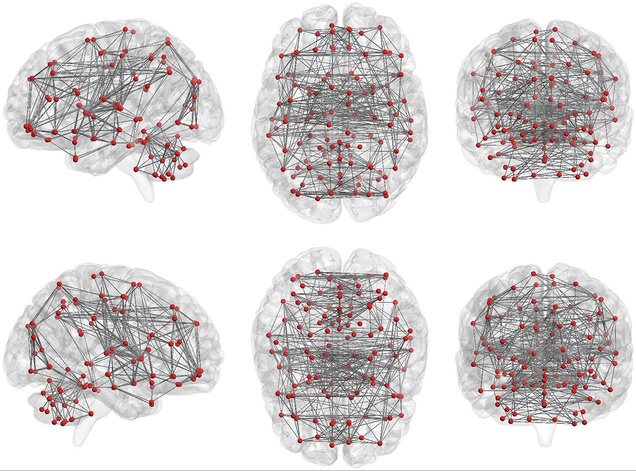
Nodes and functional connections are the basis of graph analysis. The whole brain includes 116 seeds (red dots; 45 nodes in each hemisphere of the cerebrum and 26 nodes in cerebellum) set on the basis of the anatomic parcellation defined by the Automated Anatomical Labeling atlas. Lines represent possible functional connections between those seeds.

  • 1) “Clustering coefficient” describes the level of local neighborhood clustering. It reflects the level of local connectedness of a network.

  • 2) “Characteristic path length” describes the average number of connections between all pairs of nodes. It reflects the global connectivity of the network, which reflects the efficiency of the network.

  • 3) “Node degree” describes the number of connections of a node. It helps identify the highly connected nodes within the network.

  • 4) “Centrality” describes the number of short-range connections for each node. Nodes with higher centrality contribute more to the overall efficiency of the network.

  • 5) “Modularity” describes the extent to which groups of nodes are connected to the members of their own group. It reflects the existence of subnetworks within the full network.

Graph analysis of rs-fMRI reveals a highly efficient organization of the brain network optimized toward a high level of local and global efficiency,81,82 often referred to as small-world topology. The small-world topology could characterize the whole-brain map as well as different brain networks. Graph analysis can be automatically performed using published software,88 without any a priori assumptions and with minimal bias. However, the results are often not intuitive and may be difficult to interpret.

Combined Study Recommendations

rs-fMRI has been widely used to characterize neuropsychiatric disorders using many of the methods described above.89 There are multiple methods available to identify networks within rs-fMRI data on the basis of the connectivity using data-driven parcellation and on anatomic priors. Each method emphasizes different approaches to defining brain connectivity. There is no single method currently considered a criterion standard on its own. Thus, these different methods are complementary to each other. The application of rs-fMRI is facilitated by methods that use a priori definitions of brain regions and brain networks. Combining different methods is one opportunity for yielding a more complete data-driven characterization of whole-brain resting connectivity than may be possible using one of the currently available single methods. An example of such a combination is the use of FCD, fractional-ALFF, or ReHo analysis, based on a graph theory framework, to identify ROIs for the functional connectivity analysis, as described above.

New Horizons

rs-fMRI is a rapidly evolving field with new analytic techniques introduced on a very regular basis. One such new methodology currently being proposed is the “chronnectome.”90 So far, researchers mainly focused on the temporal properties of functional connectivity between different brain regions, implicitly assuming that functional connectivity (or traffic between cities) during the scanning time is relatively static. The chronnectome approach is based on a different assumption, that of temporal evolution of spatial properties of functional connectivity (dynamic, nonstationary patterns of traffic between cities) during the scanning time. Chronnectome approaches have shown promising results for studying the spatiotemporal dynamic features of brain networks in healthy subjects91⇓⇓⇓–95 and patients.43,96⇓⇓⇓⇓–101

Conclusions

rs-fMRI is an imaging technique that plays a growing role in characterizing normal and abnormal functional brain connectivity in a variety of clinical conditions. To date, there are several different approaches and techniques to analyze rs-fMRI data, and the number of available methods is continually expanding. The different analysis methods are complementary, and applying several methods to the same dataset may yield better results compared with applying 1 method alone. A better understanding of each processing method is helpful in interpreting the common/divergent findings reported in the rs-fMRI literature.

Footnotes

  • Disclosures: Leanne M. Williams—UNRELATED: Board Membership: PsyberGuide, Comments: member of the Scientific Advisory Board; Grants/Grants Pending: National Institutes of Health*; Payment for Lectures Including Service on Speakers Bureaus: University of Minnesota, Comments: Grand Rounds. Greg Zaharchuk—UNRELATED: Grants/Grants Pending: National Institutes of Health, GE Healthcare. Michael Zeineh—UNRELATED: Grants/Grants Pending: National Institutes of Health, Doris Duke Charitable Foundation, Dana Foundation, Foundation of the ASNR, GE Healthcare*. Tali M. Ball—RELATED: Grant: National Institute of Mental Health, Comments: Salary support was provided by National Institute of Mental Health training grant T32MH019938; UNRELATED: Other: PsyberGuide, Comments: one-time payment to provide content for their Web site. Max Wintermark—UNRELATED: Board Membership: GE NFL Advisory Board. *Money paid to the institution.

  • Dr Ball is supported by grant T32MH019938 from the National Institute of Mental Health.

  • The content of this article is solely the responsibility of the authors and does not necessarily represent the official views of the National Institutes of Health.

Indicates open access to non-subscribers at www.ajnr.org

References

  1. 1.↵
    1. Handwerker DA,
    2. Bandettini PA
    . Hemodynamic signals not predicted? Not so: a comment on Sirotin and Das (2009). Neuroimage 2011;55:1409–12 doi:10.1016/j.neuroimage.2010.04.037 pmid:20406693
    CrossRefPubMed
  2. 2.↵
    1. Sirotin YB,
    2. Das A
    . Anticipatory haemodynamic signals in sensory cortex not predicted by local neuronal activity. Nature 2009;457:475–79 doi:10.1038/nature07664 pmid:19158795
    CrossRefPubMed
  3. 3.↵
    1. Nasrallah FA,
    2. Yeow LY,
    3. Biswal B, et al
    . Dependence of BOLD signal fluctuation on arterial blood CO2 and O2: implication for resting-state functional connectivity. Neuroimage 2015;117:29–39 doi:10.1016/j.neuroimage.2015.05.035 pmid:26003858
    CrossRefPubMed
  4. 4.↵
    1. Ogawa S,
    2. Lee TM,
    3. Kay AR, et al
    . Brain magnetic resonance imaging with contrast dependent on blood oxygenation. Proc Natl Acad Sci U S A 1990;87:9868–72 doi:10.1073/pnas.87.24.9868 pmid:2124706
    Abstract/FREE Full Text
  5. 5.↵
    1. Kwong KK,
    2. Belliveau JW,
    3. Chesler DA, et al
    . Dynamic magnetic resonance imaging of human brain activity during primary sensory stimulation. Proc Natl Acad Sci U S A 1992;89:5675–79 doi:10.1073/pnas.89.12.5675 pmid:1608978
    Abstract/FREE Full Text
  6. 6.↵
    1. Hyder F,
    2. Rothman DL
    . Neuronal correlate of BOLD signal fluctuations at rest: err on the side of the baseline. Proc Natl Acad Sci U S A 2010;107:10773–74 doi:10.1073/pnas.1005135107 pmid:20534504
    FREE Full Text
  7. 7.↵
    1. Schölvinck ML,
    2. Maier A,
    3. Ye FQ, et al
    . Neural basis of global resting-state fMRI activity. Proc Natl Acad Sci U S A 2010;107:10238–43 doi:10.1073/pnas.0913110107 pmid:20439733
    Abstract/FREE Full Text
  8. 8.↵
    1. Biswal B,
    2. Yetkin FZ,
    3. Haughton VM, et al
    . Functional connectivity in the motor cortex of resting human brain using echo-planar MRI. Magn Reson Med 1995;34:537–41 doi:10.1002/mrm.1910340409 pmid:8524021
    CrossRefPubMed
  9. 9.↵
    1. Smith K
    . Brain imaging: fMRI 2.0. Nature 2012;484:24–26 doi:10.1038/484024a pmid:22481337
    CrossRefPubMed
  10. 10.↵
    1. Hermes D,
    2. Nguyen M,
    3. Winawer J
    . Neuronal synchrony and the relation between the blood-oxygen-level dependent response and the local field potential. PLoS Biol 2017;15:e2001461 doi:10.1371/journal.pbio.2001461 pmid:28742093
    CrossRefPubMed
  11. 11.↵
    1. Koch C,
    2. Reid RC
    . Neuroscience: observatories of the mind. Nature 2012;483:397–98 doi:10.1038/483397a pmid:22437592
    CrossRefPubMed
  12. 12.↵
    1. Christen T,
    2. Jahanian H,
    3. Ni WW, et al
    . Noncontrast mapping of arterial delay and functional connectivity using resting-state functional MRI: a study in Moyamoya patients. J Magn Reson Imaging 2015;41:424–30 doi:10.1002/jmri.24558 pmid:24419985
    CrossRefPubMed
  13. 13.↵
    1. Gawryluk JR,
    2. Mazerolle EL,
    3. D'Arcy RC
    . Does functional MRI detect activation in white matter? A review of emerging evidence, issues, and future directions. Front Neurosci 2014;8:239 doi:10.3389/fnins.2014.00239 pmid:25152709
    CrossRefPubMed
  14. 14.↵
    1. Beissner F
    . Functional MRI of the brainstem: common problems and their solutions. Clin Neuroradiol 2015;25(Suppl 2):251–57 doi:10.1007/s00062-015-0404-0 pmid:25981409
    CrossRefPubMed
  15. 15.↵
    1. Wintermark M,
    2. Sanelli PC,
    3. Anzai Y, et al
    ; American College of Radiology Head Injury Institute. Imaging evidence and recommendations for traumatic brain injury: advanced neuro- and neurovascular imaging techniques. AJNR Am J Neuroradiol 2015;36:E1–E11 doi:10.3174/ajnr.A4181 pmid:25424870
    Abstract/FREE Full Text
  16. 16.↵
    1. Mayer AR,
    2. Bellgowan PS,
    3. Hanlon FM
    . Functional magnetic resonance imaging of mild traumatic brain injury. Neurosci Biobehav Rev 2014;49:8–18 doi:10.1016/j.neubiorev.2014.11.016 pmid:25434880
    CrossRefPubMed
  17. 17.↵
    1. Stevens MC,
    2. Lovejoy D,
    3. Kim J, et al
    . Multiple resting state network functional connectivity abnormalities in mild traumatic brain injury. Brain Imaging Behav 2012;6:293–318 doi:10.1007/s11682-012-9157-4 pmid:22555821
    CrossRefPubMed
  18. 18.↵
    1. Ishaque M,
    2. Manning JH,
    3. Woolsey MD, et al
    . Functional integrity in children with anoxic brain injury from drowning. Hum Brain Mapp 2017;38:4813–31 doi:10.1002/hbm.23745 pmid:28759710
    CrossRefPubMed
  19. 19.↵
    1. Arngrim N,
    2. Hougaard A,
    3. Schytz HW, et al
    . Effect of hypoxia on BOLD fMRI response and total cerebral blood flow in migraine with aura patients. J Cereb Blood Flow Metab 2017 Jan 1. [Epub ahead of print] doi:10.1177/0271678X17719430 pmid:28686073
    CrossRefPubMed
  20. 20.↵
    1. Lv Y,
    2. Margulies DS,
    3. Cameron CR, et al
    . Identifying the perfusion deficit in acute stroke with resting-state functional magnetic resonance imaging. Ann Neurol 2013;73:136–40 doi:10.1002/ana.23763 pmid:23378326
    CrossRefPubMed
  21. 21.↵
    1. Amemiya S,
    2. Kunimatsu A,
    3. Saito N, et al
    . Cerebral hemodynamic impairment: assessment with resting-state functional MR imaging. Radiology 2014;270:548–55 doi:10.1148/radiol.13130982 pmid:24072777
    CrossRefPubMed
  22. 22.↵
    1. Korgaonkar MS,
    2. Ram K,
    3. Williams LM, et al
    . Establishing the resting state default mode network derived from functional magnetic resonance imaging tasks as an endophenotype: a twins study. Hum Brain Mapp 2014;35:3893–902 doi:10.1002/hbm.22446 pmid:24453120
    CrossRefPubMed
  23. 23.↵
    1. Smith K
    . Brain decoding: reading minds. Nature 2013;502:428–30 doi:10.1038/502428a pmid:24153277
    CrossRefPubMed
  24. 24.↵
    1. Williams LM
    . Defining biotypes for depression and anxiety based on large-scale circuit dysfunction: a theoretical review of the evidence and future directions for clinical translation. Depress Anxiety 2017;34:9–24 doi:10.1002/da.22556 pmid:27653321
    CrossRefPubMed
  25. 25.↵
    1. Khazaee A,
    2. Ebrahimzadeh A,
    3. Babajani-Feremi A
    . Classification of patients with MCI and AD from healthy controls using directed graph measures of resting-state fMRI. Brain Behav Res 2017;322:339–50 doi:10.1016/j.bbr.2016.06.043 pmid:27345822
    CrossRefPubMed
  26. 26.↵
    1. Schirner M,
    2. Rothmeier S,
    3. Jirsa VK, et al
    . An automated pipeline for constructing personalized virtual brains from multimodal neuroimaging data. Neuroimage 2015;117:343–57 doi:10.1016/j.neuroimage.2015.03.055 pmid:25837600
    CrossRefPubMed
  27. 27.↵
    1. Chao-Gan Y,
    2. Yu-Feng Z
    . DPARSF: a MATLAB toolbox for “Pipeline” data analysis of resting-state fMRI. Front Syst Neurosci 2010;4:13 doi:10.3389/fnsys.2010.00013 pmid:20577591
    CrossRefPubMed
  28. 28.↵
    1. Dance A
    . Neuroscience: connectomes make the map. Nature 2015;526:147–49 doi:10.1038/526147a pmid:26432252
    CrossRefPubMed
  29. 29.↵
    1. Liu Y,
    2. Gao JH,
    3. Liotti M, et al
    . Temporal dissociation of parallel processing in the human subcortical outputs. Nature 1999;400:364–67 doi:10.1038/22547 pmid:10432114
    CrossRefPubMed
  30. 30.↵
    1. Tononi G,
    2. Sporns O,
    3. Edelman GM
    . A measure for brain complexity: relating functional segregation and integration in the nervous system. Proc Natl Acad Sci U S A 1994;91:5033–37 doi:10.1073/pnas.91.11.5033 pmid:8197179
    Abstract/FREE Full Text
  31. 31.↵
    1. Han L,
    2. Zhaohui L,
    3. Fei Y, et al
    . Disrupted neural activity in unilateral vascular pulsatile tinnitus patients in the early stage of disease: evidence from resting-state fMRI. Prog Neuropsychopharmacol Biol Psychiatry 2015;59:91–99 doi:10.1016/j.pnpbp.2015.01.013 pmid:25645870
    CrossRefPubMed
  32. 32.↵
    1. Premi E,
    2. Cauda F,
    3. Gasparotti R, et al
    . Multimodal FMRI resting-state functional connectivity in granulin mutations: the case of fronto-parietal dementia. PLoS One 2014;9:e106500 doi:10.1371/journal.pone.0106500 pmid:25188321
    CrossRefPubMed
  33. 33.↵
    1. An L,
    2. Cao QJ,
    3. Sui MQ, et al
    . Local synchronization and amplitude of the fluctuation of spontaneous brain activity in attention-deficit/hyperactivity disorder: a resting-state fMRI study. Neurosci Bull 2013;29:603–13 doi:10.1007/s12264-013-1353-8 pmid:23861089
    CrossRefPubMed
  34. 34.↵
    1. Lei D,
    2. Ma J,
    3. Du X, et al
    . Spontaneous brain activity changes in children with primary monosymptomatic nocturnal enuresis: a resting-state fMRI study. Neurourol Urodyn 2012;31:99–104 doi:10.1002/nau.21205 pmid:22038619
    CrossRefPubMed
  35. 35.↵
    1. Zang YF,
    2. He Y,
    3. Zhu CZ, et al
    . Altered baseline brain activity in children with ADHD revealed by resting-state functional MRI. Brain Dev 2007;29:83–91 doi:10.1016/j.braindev.2006.07.002 pmid:16919409
    CrossRefPubMed
  36. 36.↵
    1. Song XW,
    2. Dong ZY,
    3. Long XY, et al
    . REST: a toolkit for resting-state functional magnetic resonance imaging data processing. PLoS One 2011;6:e25031 doi:10.1371/journal.pone.0025031 pmid:21949842
    CrossRefPubMed
  37. 37.↵
    1. Zou QH,
    2. Zhu CZ,
    3. Yang Y, et al
    . An improved approach to detection of Amplitude of Low-Frequency Fluctuation (ALFF) for resting-state fMRI: fractional ALFF. J Neurosci Methods 2008;172:137–41 doi:10.1016/j.jneumeth.2008.04.012 pmid:18501969
    CrossRefPubMed
  38. 38.↵
    1. Buzsáki G,
    2. Draguhn A
    . Neuronal oscillations in cortical networks. Science 2004;304:1926–29 doi:10.1126/science.1099745 pmid:15218136
    Abstract/FREE Full Text
  39. 39.↵
    1. Zuo XN,
    2. Di Martino A,
    3. Kelly C, et al
    . The oscillating brain: complex and reliable. Neuroimage 2010;49:1432–45 doi:10.1016/j.neuroimage.2009.09.037 pmid:19782143
    CrossRefPubMed
  40. 40.↵
    1. Xue SW,
    2. Li D,
    3. Weng XC, et al
    . Different neural manifestations of two slow frequency bands in resting functional magnetic resonance imaging: a systemic survey at regional, interregional, and network levels. Brain Connect 2014;4:242–55 doi:10.1089/brain.2013.0182 pmid:24456196
    CrossRefPubMed
  41. 41.↵
    1. Liu X,
    2. Wang S,
    3. Zhang X, et al
    . Abnormal amplitude of low-frequency fluctuations of intrinsic brain activity in Alzheimer's disease. J Alzheimers Dis 2014;40:387–97 doi:10.3233/JAD-131322 pmid:24473186
    CrossRefPubMed
  42. 42.↵
    1. Xue S,
    2. Wang X,
    3. Wang W, et al
    . Frequency-dependent alterations in regional homogeneity in major depression. Brain Behav Res 2016;306:13–19 doi:10.1016/j.bbr.2016.03.012 pmid:26968135
    CrossRefPubMed
  43. 43.↵
    1. Chen X,
    2. Zhang H,
    3. Zhang L, et al
    . Extraction of dynamic functional connectivity from brain grey matter and white matter for MCI classification. Hum Brain Mapp 2017;38:5019–34 doi:10.1002/hbm.23711 pmid:28665045
    CrossRefPubMed
  44. 44.↵
    1. Zhang L,
    2. Zhang H,
    3. Chen X, et al
    . Learning-based structurally-guided construction of resting-state functional correlation tensors. Magn Reson Imaging 2017;43:110–21 doi:10.1016/j.mri.2017.07.008 pmid:28729016
    CrossRefPubMed
  45. 45.↵
    1. Küblböck M,
    2. Woletz M,
    3. Höflich A, et al
    . Stability of low-frequency fluctuation amplitudes in prolonged resting-state fMRI. Neuroimage 2014;103:249–57 doi:10.1016/j.neuroimage.2014.09.038 pmid:25251869
    CrossRefPubMed
  46. 46.↵
    1. Zuo XN,
    2. Xing XX
    . Test-retest reliabilities of resting-state FMRI measurements in human brain functional connectomics: a systems neuroscience perspective. Neurosci Biobehav Rev 2014;45:100–18 doi:10.1016/j.neubiorev.2014.05.009 pmid:24875392
    CrossRefPubMed
  47. 47.↵
    1. Zang Y,
    2. Jiang T,
    3. Lu Y, et al
    . Regional homogeneity approach to fMRI data analysis. Neuroimage 2004;22:394–400 doi:10.1016/j.neuroimage.2003.12.030 pmid:15110032
    CrossRefPubMed
  48. 48.↵
    1. Zang YF,
    2. Zuo XN,
    3. Milham M, et al
    . Toward a meta-analytic synthesis of the resting-state fMRI literature for clinical populations. Biomed Res Int 2015;2015:435265 doi:10.1155/2015/435265 pmid:26171391
    CrossRefPubMed
  49. 49.↵
    1. Song X,
    2. Zhang Y,
    3. Liu Y
    . Frequency specificity of regional homogeneity in the resting-state human brain. PLoS One 2014;9:e86818 doi:10.1371/journal.pone.0086818 pmid:24466256
    CrossRefPubMed
  50. 50.↵
    1. Liu ZR,
    2. Miao HH,
    3. Yu Y, et al
    . Frequency-specific local synchronization changes in paroxysmal kinesigenic dyskinesia. Medicine (Baltimore) 2016;95:e3293 doi:10.1097/MD.0000000000003293 pmid:27043701
    CrossRefPubMed
  51. 51.↵
    1. Huang Z,
    2. Wang Z,
    3. Zhang J, et al
    . Altered temporal variance and neural synchronization of spontaneous brain activity in anesthesia. Hum Brain Mapp 2014;35:5368–78 doi:10.1002/hbm.22556 pmid:24867379
    CrossRefPubMed
  52. 52.↵
    1. Yu R,
    2. Hsieh MH,
    3. Wang HL, et al
    . Frequency dependent alterations in regional homogeneity of baseline brain activity in schizophrenia. PLoS One 2013;8:e57516 doi:10.1371/journal.pone.0057516 pmid:23483911
    CrossRefPubMed
  53. 53.↵
    1. Zuo XN,
    2. Xu T,
    3. Jiang L, et al
    . Toward reliable characterization of functional homogeneity in the human brain: preprocessing, scan duration, imaging resolution and computational space. Neuroimage 2013;65:374–86 doi:10.1016/j.neuroimage.2012.10.017 pmid:23085497
    CrossRefPubMed
  54. 54.↵
    1. Cui Y,
    2. Jiao Y,
    3. Chen YC, et al
    . Altered spontaneous brain activity in type 2 diabetes: a resting-state functional MRI study. Diabetes 2014;63:749–60 doi:10.2337/db13-0519 pmid:24353185
    Abstract/FREE Full Text
  55. 55.↵
    1. Premi E,
    2. Cauda F,
    3. Gasparotti R, et al
    . Multimodal fMRI resting-state functional connectivity in granulin mutations: the case of fronto-parietal dementia. PLoS One 2014;9:e106500 doi:10.1371/journal.pone.0106500 pmid:25188321
    CrossRefPubMed
  56. 56.↵
    1. Lanting CP,
    2. de Kleine E,
    3. Langers DR, et al
    . Unilateral tinnitus: changes in connectivity and response lateralization measured with fMRI. PLoS One 2014;9:e110704 doi:10.1371/journal.pone.0110704 pmid:25329557
    CrossRefPubMed
  57. 57.↵
    1. Friston KJ
    . Modalities, modes, and models in functional neuroimaging. Science 2009;326:399–403 doi:10.1126/science.1174521 pmid:19833961
    Abstract/FREE Full Text
  58. 58.↵
    1. Tomasi D,
    2. Volkow ND
    . Functional connectivity density mapping. Proc Natl Acad Sci U S A 2010;107:9885–90 doi:10.1073/pnas.1001414107 pmid:20457896
    Abstract/FREE Full Text
  59. 59.↵
    1. Achard S,
    2. Salvador R,
    3. Whitcher B, et al
    . A resilient, low-frequency, small-world human brain functional network with highly connected association cortical hubs. J Neurosci 2006;26:63–72 doi:10.1523/JNEUROSCI.3874-05.2006 pmid:16399673
    Abstract/FREE Full Text
  60. 60.↵
    1. He Y,
    2. Chen ZJ,
    3. Evans AC
    . Small-world anatomical networks in the human brain revealed by cortical thickness from MRI. Cereb Cortex 2007;17:2407–19 doi:10.1093/cercor/bhl149 pmid:17204824
    CrossRefPubMed
  61. 61.↵
    1. Xia M,
    2. Wang J,
    3. He Y
    . BrainNet Viewer: a network visualization tool for human brain connectomics. PLoS One 2013;8:e68910 doi:10.1371/journal.pone.0068910 pmid:23861951
    CrossRefPubMed
  62. 62.↵
    1. Lee MH,
    2. Smyser CD,
    3. Shimony JS
    . Resting-state fMRI: a review of methods and clinical applications. AJNR Am J Neuroradiol 2013;34:1866–72 doi:10.3174/ajnr.A3263 pmid:22936095
    Abstract/FREE Full Text
  63. 63.↵
    1. Wilson PH,
    2. Smits-Engelsman B,
    3. Caeyenberghs K, et al
    . Cognitive and neuroimaging findings in developmental coordination disorder: new insights from a systematic review of recent research. Dev Med Child Neurol 2017;59:1117–29 doi:10.1111/dmcn.13530 pmid:28872667
    CrossRefPubMed
  64. 64.↵
    1. Koyama MS,
    2. Parvaz MA,
    3. Goldstein RZ
    . The adolescent brain at risk for substance use disorders: a review of functional MRI research on motor response inhibition. Curr Opin Behav Sci 2017;13:186–95 doi:10.1016/j.cobeha.2016.12.006 pmid:28868337
    CrossRefPubMed
  65. 65.↵
    1. Gravel N,
    2. Harvey BM,
    3. Renken RJ, et al
    . Phase-synchronization-based parcellation of resting state fMRI signals reveals topographically organized clusters in early visual cortex. Neuroimage 2017 Sep 1. [Epub ahead of print] doi:10.1016/j.neuroimage.2017.08.063 pmid:28867341
    CrossRefPubMed
  66. 66.↵
    1. Kiviniemi V,
    2. Kantola JH,
    3. Jauhiainen J, et al
    . Independent component analysis of nondeterministic fMRI signal sources. Neuroimage 2003;19:253–60 doi:10.1016/S1053-8119(03)00097-1 pmid:12814576
    CrossRefPubMed
  67. 67.↵
    1. van de Ven VG,
    2. Formisano E,
    3. Prvulovic D, et al
    . Functional connectivity as revealed by spatial independent component analysis of fMRI measurements during rest. Hum Brain Mapp 2004;22:165–78 doi:10.1002/hbm.20022 pmid:15195284
    CrossRefPubMed
  68. 68.↵
    1. Beckmann CF,
    2. DeLuca M,
    3. Devlin JT, et al
    . Investigations into resting-state connectivity using independent component analysis. Philos Trans R Soc Lond B Biol Sci 2005;360:1001–13 doi:10.1098/rstb.2005.1634 pmid:16087444
    Abstract/FREE Full Text
  69. 69.↵
    1. Shirer WR,
    2. Ryali S,
    3. Rykhlevskaia E, et al
    . Decoding subject-driven cognitive states with whole-brain connectivity patterns. Cereb Cortex 2012;22:158–65 doi:10.1093/cercor/bhr099 pmid:21616982
    CrossRefPubMed
  70. 70.↵
    1. Seeley WW,
    2. Menon V,
    3. Schatzberg AF, et al
    . Dissociable intrinsic connectivity networks for salience processing and executive control. J Neurosci 2007;27:2349–56 doi:10.1523/JNEUROSCI.5587-06.2007 pmid:17329432
    Abstract/FREE Full Text
  71. 71.↵
    1. Zuo XN,
    2. Kelly C,
    3. Adelstein JS, et al
    . Reliable intrinsic connectivity networks: test-retest evaluation using ICA and dual regression approach. Neuroimage 2010;49:2163–77 doi:10.1016/j.neuroimage.2009.10.080 pmid:19896537
    CrossRefPubMed
  72. 72.↵
    1. Bertolero MA,
    2. Yeo BT,
    3. D'Esposito M
    . The modular and integrative functional architecture of the human brain. Proc Natl Acad Sci U S A 2015;112:E6798–807 doi:10.1073/pnas.1510619112 pmid:26598686
    Abstract/FREE Full Text
  73. 73.↵
    1. Anderson M,
    2. Fu GS,
    3. Phlypo R, et al
    . Independent vector analysis: identification conditions and performance bounds. IEEE Trans Signal Process 2014;62:4399–410 doi:10.1109/TSP.2014.2333554
    CrossRef
  74. 74.↵
    1. Lee JH,
    2. Lee TW,
    3. Jolesz FA, et al
    . Independent vector analysis (IVA): multivariate approach for fMRI group study. Neuroimage 2008;40:86–109 doi:10.1016/j.neuroimage.2007.11.019 pmid:18165105
    CrossRefPubMed
  75. 75.↵
    1. Anderson M,
    2. Adali T,
    3. Li XL
    . Joint blind source separation with multivariate Gaussian model: algorithms and performance analysis. IEEE Trans Signal Process 2012;60:1672–83
  76. 76.↵
    1. Laney J,
    2. Westlake KP,
    3. Ma S, et al
    . Capturing subject variability in fMRI data: a graph-theoretical analysis of GICA vs. IVA. J Neurosci Methods 2015;247:32–40 doi:10.1016/j.jneumeth.2015.03.019 pmid:25797843
    CrossRefPubMed
  77. 77.↵
    1. Ma S,
    2. Phlypo R,
    3. Calhoun VD, et al
    . Capturing group variability using IVA: a simulation study and graph-theoretical analysis. Proc IEEE Int Conf Acoust Speech Signal Process 2013:3128–32 doi:10.1109/ICASSP.2013.6638234
    CrossRef
  78. 78.↵
    1. Allen EA,
    2. Erhardt EB,
    3. Wei Y, et al
    . Capturing inter-subject variability with group independent component analysis of fMRI data: a simulation study. Neuroimage 2012;59:4141–59 doi:10.1016/j.neuroimage.2011.10.010 pmid:22019879
    CrossRefPubMed
  79. 79.↵
    1. Watts DJ,
    2. Strogatz SH
    . Collective dynamics of ‘small-world’ networks. Nature 1998;393:440–42 doi:10.1038/30918 pmid:9623998
    CrossRefPubMed
  80. 80.↵
    1. Liao X,
    2. Vasilakos AV,
    3. He Y
    . Small-world human brain networks: perspectives and challenges. Neurosci Biobehav Rev 2017;77:286–300 doi:10.1016/j.neubiorev.2017.03.018 pmid:28389343
    CrossRefPubMed
  81. 81.↵
    1. Bullmore E,
    2. Sporns O
    . Complex brain networks: graph theoretical analysis of structural and functional systems. Nat Rev Neurosci 2009;10:186–98 doi:10.1038/nrn2575 pmid:19190637
    CrossRefPubMed
  82. 82.↵
    1. Sporns O,
    2. Honey CJ,
    3. Kotter R
    . Identification and classification of hubs in brain networks. PLoS One 2007;2:e1049 doi:10.1371/journal.pone.0001049 pmid:17940613
    CrossRefPubMed
  83. 83.↵
    1. van den Heuvel MP,
    2. Stam CJ,
    3. Boersma M, et al
    . Small-world and scale-free organization of voxel-based resting-state functional connectivity in the human brain. Neuroimage 2008;43:528–39 doi:10.1016/j.neuroimage.2008.08.010 pmid:18786642
    CrossRefPubMed
  84. 84.↵
    1. Achard S,
    2. Bullmore E
    . Efficiency and cost of economical brain functional networks. PLoS Comput Biol 2007;3:e17 doi:10.1371/journal.pcbi.0030017 pmid:17274684
    CrossRefPubMed
  85. 85.↵
    1. Glasser MF,
    2. Coalson TS,
    3. Robinson EC, et al
    . A multi-modal parcellation of human cerebral cortex. Nature 2016;536:171–78 doi:10.1038/nature18933 pmid:27437579
    CrossRefPubMed
  86. 86.↵
    1. Zalesky A,
    2. Fornito A,
    3. Harding IH, et al
    . Whole-brain anatomical networks: does the choice of nodes matter? Neuroimage 2010;50:970–83 doi:10.1016/j.neuroimage.2009.12.027 pmid:20035887
    CrossRefPubMed
  87. 87.↵
    1. Li W,
    2. Andreasen NC,
    3. Nopoulos P, et al
    . Automated parcellation of the brain surface generated from magnetic resonance images. Front Neuroinform 2013;7:23 doi:10.3389/fninf.2013.00023 pmid:24155718
    CrossRefPubMed
  88. 88.↵
    1. Wang J,
    2. Wang X,
    3. Xia M, et al
    . GRETNA: a graph theoretical network analysis toolbox for imaging connectomics. Front Hum Neurosci 2015;9:386 doi:10.3389/fnhum.2015.00386 pmid:26175682
    CrossRefPubMed
  89. 89.↵
    1. Menon V
    . Large-scale brain networks and psychopathology: a unifying triple network model. Trends Cogn Sci 2011;15:483–506 doi:10.1016/j.tics.2011.08.003 pmid:21908230
    CrossRefPubMed
  90. 90.↵
    1. Calhoun VD,
    2. Miller R,
    3. Pearlson G, et al
    . The chronnectome: time-varying connectivity networks as the next frontier in fMRI data discovery. Neuron 2014;84:262–74 doi:10.1016/j.neuron.2014.10.015 pmid:25374354
    CrossRefPubMed
  91. 91.↵
    1. Griffa A,
    2. Ricaud B,
    3. Benzi K, et al
    . Transient networks of spatio-temporal connectivity map communication pathways in brain functional systems. Neuroimage 2017;155:490–502 doi:10.1016/j.neuroimage.2017.04.015 pmid:28412440
    CrossRefPubMed
  92. 92.↵
    1. Abrol A,
    2. Damaraju E,
    3. Miller RL, et al
    . Replicability of time-varying connectivity patterns in large resting state fMRI samples. Neuroimage 2017;163:160–76 doi:10.1016/j.neuroimage.2017.09.020 pmid:28916181
    CrossRefPubMed
  93. 93.↵
    1. Abrol A,
    2. Chaze C,
    3. Damaraju E, et al
    . The chronnectome: evaluating replicability of dynamic connectivity patterns in 7500 resting fMRI datasets. Conf Proc IEEE Eng Med Biol Soc 2016;2016:5571–74 doi:10.1109/EMBC.2016.7591989 pmid:28269517
    CrossRefPubMed
  94. 94.↵
    1. Thompson WH,
    2. Fransson P
    . The frequency dimension of fMRI dynamic connectivity: network connectivity, functional hubs and integration in the resting brain. Neuroimage 2015;121:227–42 doi:10.1016/j.neuroimage.2015.07.022 pmid:26169321
    CrossRefPubMed
  95. 95.↵
    1. Chen Y,
    2. Wang W,
    3. Zhao X, et al
    . Age-related decline in the variation of dynamic functional connectivity: a resting state analysis. Front Aging Neurosci 2017;9:203 doi:10.3389/fnagi.2017.00203 pmid:28713261
    CrossRefPubMed
  96. 96.↵
    1. Park BY,
    2. Moon T,
    3. Park H
    . Dynamic functional connectivity analysis reveals improved association between brain networks and eating behaviors compared to static analysis. Brain Behav Res 2018;337:114–21 doi:10.1016/j.bbr.2017.10.001 pmid:28986105
    CrossRefPubMed
  97. 97.↵
    1. Cheng JC,
    2. Bosma RL,
    3. Hemington KS, et al
    . Slow-5 dynamic functional connectivity reflects the capacity to sustain cognitive performance during pain. Neuroimage 2017;157:61–68 doi:10.1016/j.neuroimage.2017.06.005 pmid:28583880
    CrossRefPubMed
  98. 98.↵
    1. Damaraju E,
    2. Allen EA,
    3. Belger A, et al
    . Dynamic functional connectivity analysis reveals transient states of dysconnectivity in schizophrenia. Neuroimage Clin 2014;5:298–308 doi:10.1016/j.nicl.2014.07.003 pmid:25161896
    CrossRefPubMed
  99. 99.↵
    1. Hutchison RM,
    2. Womelsdorf T,
    3. Allen EA, et al
    . Dynamic functional connectivity: promise, issues, and interpretations. Neuroimage 2013;80:360–78 doi:10.1016/j.neuroimage.2013.05.079 pmid:23707587
    CrossRefPubMed
  100. 100.↵
    1. Preti MG,
    2. Bolton TA,
    3. Van De Ville D
    . The dynamic functional connectome: state-of-the-art and perspectives. Neuroimage 2017;160:41–54 doi:10.1016/j.neuroimage.2016.12.061 pmid:28034766
    CrossRefPubMed
  101. 101.↵
    1. Lindquist MA,
    2. Xu Y,
    3. Nebel MB, et al
    . Evaluating dynamic bivariate correlations in resting-state fMRI: a comparison study and a new approach. Neuroimage 2014;101:531–46 doi:10.1016/j.neuroimage.2014.06.052 pmid:24993894
    CrossRefPubMed
  • © 2018 by American Journal of Neuroradiology
View Abstract
Next
Back to top
Advertisement
Print
Download PDF
Email Article

Thank you for your interest in spreading the word on American Journal of Neuroradiology.

NOTE: We only request your email address so that the person you are recommending the page to knows that you wanted them to see it, and that it is not junk mail. We do not capture any email address.

Enter multiple addresses on separate lines or separate them with commas.
Resting-State Functional MRI: Everything That Nonexperts Have Always Wanted to Know
(Your Name) has sent you a message from American Journal of Neuroradiology
(Your Name) thought you would like to see the American Journal of Neuroradiology web site.
CAPTCHA
This question is for testing whether or not you are a human visitor and to prevent automated spam submissions.
Cite this article
H. Lv, Z. Wang, E. Tong, L.M. Williams, G. Zaharchuk, M. Zeineh, A.N. Goldstein-Piekarski, T.M. Ball, C. Liao, M. Wintermark
Resting-State Functional MRI: Everything That Nonexperts Have Always Wanted to Know
American Journal of Neuroradiology Jan 2018, DOI: 10.3174/ajnr.A5527

Citation Manager Formats

  • BibTeX
  • Bookends
  • EasyBib
  • EndNote (tagged)
  • EndNote 8 (xml)
  • Medlars
  • Mendeley
  • Papers
  • RefWorks Tagged
  • Ref Manager
  • RIS
  • Zotero
0 Responses
Respond to this article
Share
Bookmark this article
Resting-State Functional MRI: Everything That Nonexperts Have Always Wanted to Know
H. Lv, Z. Wang, E. Tong, L.M. Williams, G. Zaharchuk, M. Zeineh, A.N. Goldstein-Piekarski, T.M. Ball, C. Liao, M. Wintermark
American Journal of Neuroradiology Jan 2018, DOI: 10.3174/ajnr.A5527
del.icio.us logo Twitter logo Facebook logo Mendeley logo
  • Tweet Widget
  • Facebook Like
  • Google Plus One
Purchase

Jump to section

  • Article
    • Abstract
    • ABBREVIATIONS:
    • Conclusions
    • Footnotes
    • References
  • Figures & Data
  • Info & Metrics
  • Responses
  • References
  • PDF

Related Articles

  • No related articles found.
  • PubMed
  • Google Scholar

Cited By...

  • Exploring dynamic alpha band connectivity in Parkinsons disease: A novel approach to postural control assessment using the BioVRSea paradigm
  • Cognitive reserve linked to network-specific brain-ventricle coupling
  • Apparent Diffusion Coefficient fMRI shines light on white matter resting-state connectivity compared to BOLD
  • Within-Individual Precision Mapping of Brain Networks Exclusively Using Task Data
  • How does methamphetamine affect the brain? A systematic review of magnetic resonance imaging studies
  • Genetic and environmental contributions to ReHo and fALFF in early adolescence vary across brain regions
  • Evidence for white matter intrinsic connectivity networks at rest and during a task: a large-scale study and templates
  • Addressing Inconsistency in Functional Neuroimaging: A Replicable Data-Driven Multi-Scale Functional Atlas for Canonical Brain Networks
  • Noradrenergic and Dopaminergic Neural Correlates of Trait Anxiety: Unveiling the Impact of Maladaptive Emotion Regulation
  • Functional Activity, Functional Connectivity and Complex Network Biomarkers of Progressive Hyposmia Parkinsons Disease with No Cognitive Impairment: Evidences from Resting-state fMRI Study
  • A Comprehensive and Broad Approach to Resting-State Functional Connectivity in Adult Patients with Mild Traumatic Brain Injury
  • Co-representation of Functional Brain Networks Is Shaped by Cortical Myeloarchitecture and Reveals Individual Behavioral Ability
  • A Cross-Sectional and Longitudinal Integrated Study on Brain Functional Changes in a Neuropathic Pain Rat Model
  • Corticospinal excitability at rest outside of a task does not differ from task intertrial intervals in healthy adults
  • Priming transcranial direct current stimulation for improving hemiparetic upper limb in patients with subacute stroke: study protocol for a randomised controlled trial
  • Right-to-Left Shunt-associated Brain Functional Changes in Migraine: Evidences from a Resting-state FMRI Study
  • Brain connectivity changes underlying depression and fatigue in relapsing-remitting multiple sclerosis: a systematic review
  • Connectivity Regression
  • Genetic influences on the intrinsic and extrinsic functional organizations of the cerebral cortex
  • N-acetyl Cysteine attenuates intrinsic functional connectivity, but not neural alcohol cue reactivity, in treatment-seeking individuals with alcohol use disorder
  • Prosocial Decision-Making and Human Brain: A Graph Theory Analysis on Resting-State Functional Magnetic Resonance Image
  • Seed-based morphometry of nodes in the default mode network among patients with Alzheimers disease in Klang Valley, Malaysia
  • Alteration of functional connectivity in the cortex and major brain networks of non-human primates following focused ultrasound exposure
  • Altered regional homogeneity and its association with cognitive function in adolescents with borderline personality disorder
  • Novel inductively-coupled ear-bars (ICEs) for fMRI signal enhancement in rat entorhinal cortex
  • Assessing The Repeatability of Multi-Frequency Multi-Layer Brain Network Topologies Across Alternative Researchers Choice Paths
  • Effects of a single-dose methylphenidate challenge on resting-state functional connectivity in stimulant-treatment naive children and adults with ADHD
  • A novel rhesus macaque model of Huntingtons disease recapitulates key neuropathological changes along with progressive motor and cognitive decline
  • Chemogenetic activation of Locus Coeruleus Noradrenergic Neurons Modulates the Default Mode Network
  • Resting-state functional reorganisation in Alzheimers disease and amnestic mild cognitive impairment: protocol for a systematic review and meta-analysis
  • Resting-State Functional MRI for Determining Language Lateralization in Children with Drug-Resistant Epilepsy
  • Sex Differences in Brain Structures and Functional Connectivity among Patients with Bipolar I Disorder
  • Time-varying Dynamic Network Model For Dynamic Resting State Functional Connectivity in fMRI and MEG imaging
  • Functional network dynamics in progressive multiple sclerosis
  • Common variants contribute to intrinsic human brain functional networks
  • Structural and functional brain parameters related to cognitive performance across development: Replication and extension of the parieto-frontal integration theory in a single sample
  • Default mode network anatomy and function is linked to pediatric concussion recovery
  • Network-wide abnormalities explain memory variability in hippocampal amnesia
  • Interaction of Developmental Venous Anomalies with Resting-State Functional MRI Measures
  • Crossref (162)
  • Google Scholar

This article has been cited by the following articles in journals that are participating in Crossref Cited-by Linking.

  • Alterations of White Matter Integrity and Hippocampal Functional Connectivity in Type 2 Diabetes Without Mild Cognitive Impairment
    Qian Sun, Guan-Qun Chen, Xi-Bin Wang, Ying Yu, Yu-Chuan Hu, Lin-Feng Yan, Xin Zhang, Yang Yang, Jin Zhang, Bin Liu, Cong-Cong Wang, Yi Ma, Wen Wang, Ying Han, Guang-Bin Cui
    Frontiers in Neuroanatomy 2018 12
  • Resting-state functional connectivity predictors of treatment response in schizophrenia – A systematic review and meta-analysis
    Urvakhsh Meherwan Mehta, Ferose Azeez Ibrahim, Manu S. Sharma, Ganesan Venkatasubramanian, Jagadisha Thirthalli, Rose Dawn Bharath, Nicolas R. Bolo, Bangalore N. Gangadhar, Matcheri S. Keshavan
    Schizophrenia Research 2021 237
  • Why is Clinical fMRI in a Resting State?
    Erin E. O'Connor, Thomas A. Zeffiro
    Frontiers in Neurology 2019 10
  • Reduced resting-state functional connectivity of the basolateral amygdala to the medial prefrontal cortex in preweaning rats exposed to chronic early-life stress
    Angela Guadagno, Min Su Kang, Gabriel A. Devenyi, Axel P. Mathieu, Pedro Rosa-Neto, Mallar Chakravarty, Claire-Dominique Walker
    Brain Structure and Function 2018 223 8
  • Functional connectome of brainstem nuclei involved in autonomic, limbic, pain and sensory processing in living humans from 7 Tesla resting state fMRI
    Simone Cauzzo, Kavita Singh, Matthew Stauder, María Guadalupe García-Gomar, Nicola Vanello, Claudio Passino, Jeffrey Staab, Iole Indovina, Marta Bianciardi
    NeuroImage 2022 250
  • Electrophysiological and Neuroimaging Studies – During Resting State and Sensory Stimulation in Disorders of Consciousness: A Review
    Ritika Jain, Angarai Ganesan Ramakrishnan
    Frontiers in Neuroscience 2020 14
  • Resting-State Brain Network Dysfunctions Associated With Visuomotor Impairments in Autism Spectrum Disorder
    Zheng Wang, Yan Wang, John A. Sweeney, Qiyong Gong, Su Lui, Matthew W. Mosconi
    Frontiers in Integrative Neuroscience 2019 13
  • Network-wide abnormalities explain memory variability in hippocampal amnesia
    Georgios PD Argyropoulos, Clare Loane, Adriana Roca-Fernandez, Carmen Lage-Martinez, Oana Gurau, Sarosh R Irani, Christopher R Butler
    eLife 2019 8
  • Task-related brain functional network reconfigurations relate to motor recovery in chronic subcortical stroke
    Hsiao-Ju Cheng, Kwun Kei Ng, Xing Qian, Fang Ji, Zhong Kang Lu, Wei Peng Teo, Xin Hong, Fatima Ali Nasrallah, Kai Keng Ang, Kai-Hsiang Chuang, Cuntai Guan, Haoyong Yu, Effie Chew, Juan Helen Zhou
    Scientific Reports 2021 11 1
  • Structural and Functional Brain Parameters Related to Cognitive Performance Across Development: Replication and Extension of the Parieto-Frontal Integration Theory in a Single Sample
    Ruben C Gur, Ellyn R Butler, Tyler M Moore, Adon F G Rosen, Kosha Ruparel, Theodore D Satterthwaite, David R Roalf, Efstathios D Gennatas, Warren B Bilker, Russell T Shinohara, Allison Port, Mark A Elliott, Ragini Verma, Christos Davatzikos, Daniel H Wolf, John A Detre, Raquel E Gur
    Cerebral Cortex 2021 31 3

More in this TOC Section

  • An Atlas of Neonatal Neurovascular Imaging Anatomy as Depicted with Microvascular Imaging: The Intracranial Arteries
  • An Atlas of Neonatal Neurovascular Imaging Anatomy as Depicted with Microvascular Imaging: The Intracranial Veins
  • Clinical Translation of Hyperpolarized 13C Metabolic Probes for Glioma Imaging
Show more Review Article

Similar Articles

Advertisement

Indexed Content

  • Current Issue
  • Accepted Manuscripts
  • Article Preview
  • Past Issues
  • Editorials
  • Editor's Choice
  • Fellows' Journal Club
  • Letters to the Editor
  • Video Articles

Cases

  • Case Collection
  • Archive - Case of the Week
  • Archive - Case of the Month
  • Archive - Classic Case

Special Collections

  • AJNR Awards
  • ASNR Foundation Special Collection
  • Most Impactful AJNR Articles
  • Photon-Counting CT
  • Spinal CSF Leak Articles (Jan 2020-June 2024)

More from AJNR

  • Trainee Corner
  • Imaging Protocols
  • MRI Safety Corner

Multimedia

  • AJNR Podcasts
  • AJNR Scantastics

Resources

  • Turnaround Time
  • Submit a Manuscript
  • Submit a Video Article
  • Submit an eLetter to the Editor/Response
  • Manuscript Submission Guidelines
  • Statistical Tips
  • Fast Publishing of Accepted Manuscripts
  • Graphical Abstract Preparation
  • Imaging Protocol Submission
  • Evidence-Based Medicine Level Guide
  • Publishing Checklists
  • Author Policies
  • Become a Reviewer/Academy of Reviewers
  • News and Updates

About Us

  • About AJNR
  • Editorial Board
  • Editorial Board Alumni
  • Alerts
  • Permissions
  • Not an AJNR Subscriber? Join Now
  • Advertise with Us
  • Librarian Resources
  • Feedback
  • Terms and Conditions
  • AJNR Editorial Board Alumni

American Society of Neuroradiology

  • Not an ASNR Member? Join Now

© 2025 by the American Society of Neuroradiology All rights, including for text and data mining, AI training, and similar technologies, are reserved.
Print ISSN: 0195-6108 Online ISSN: 1936-959X

Powered by HighWire