Advertisement

Main menu

  • Home
  • Content
    • Current Issue
    • Accepted Manuscripts
    • Article Preview
    • Past Issue Archive
    • Video Articles
    • AJNR Case Collection
    • Case of the Week Archive
    • Case of the Month Archive
    • Classic Case Archive
  • Special Collections
    • AJNR Awards
    • ASNR Foundation Special Collection
    • Most Impactful AJNR Articles
    • Photon-Counting CT
    • Spinal CSF Leak Articles (Jan 2020-June 2024)
  • Multimedia
    • AJNR Podcasts
    • AJNR SCANtastic
    • Trainee Corner
    • MRI Safety Corner
    • Imaging Protocols
  • For Authors
    • Submit a Manuscript
    • Submit a Video Article
    • Submit an eLetter to the Editor/Response
    • Manuscript Submission Guidelines
    • Statistical Tips
    • Fast Publishing of Accepted Manuscripts
    • Graphical Abstract Preparation
    • Imaging Protocol Submission
    • Author Policies
  • About Us
    • About AJNR
    • Editorial Board
    • Editorial Board Alumni
  • More
    • Become a Reviewer/Academy of Reviewers
    • Subscribers
    • Permissions
    • Alerts
    • Feedback
    • Advertisers
    • ASNR Home

User menu

  • Alerts
  • Log in

Search

  • Advanced search
American Journal of Neuroradiology

American Journal of Neuroradiology

ASHNR American Society of Functional Neuroradiology ASHNR American Society of Pediatric Neuroradiology ASSR
  • Alerts
  • Log in

Advanced Search

  • Home
  • Content
    • Current Issue
    • Accepted Manuscripts
    • Article Preview
    • Past Issue Archive
    • Video Articles
    • AJNR Case Collection
    • Case of the Week Archive
    • Case of the Month Archive
    • Classic Case Archive
  • Special Collections
    • AJNR Awards
    • ASNR Foundation Special Collection
    • Most Impactful AJNR Articles
    • Photon-Counting CT
    • Spinal CSF Leak Articles (Jan 2020-June 2024)
  • Multimedia
    • AJNR Podcasts
    • AJNR SCANtastic
    • Trainee Corner
    • MRI Safety Corner
    • Imaging Protocols
  • For Authors
    • Submit a Manuscript
    • Submit a Video Article
    • Submit an eLetter to the Editor/Response
    • Manuscript Submission Guidelines
    • Statistical Tips
    • Fast Publishing of Accepted Manuscripts
    • Graphical Abstract Preparation
    • Imaging Protocol Submission
    • Author Policies
  • About Us
    • About AJNR
    • Editorial Board
    • Editorial Board Alumni
  • More
    • Become a Reviewer/Academy of Reviewers
    • Subscribers
    • Permissions
    • Alerts
    • Feedback
    • Advertisers
    • ASNR Home
  • Follow AJNR on Twitter
  • Visit AJNR on Facebook
  • Follow AJNR on Instagram
  • Join AJNR on LinkedIn
  • RSS Feeds

AJNR is seeking candidates for the AJNR Podcast Editor. Read the position description.

OtherBRAIN

Practical Visualization of Internal Structure of White Matter for Image Interpretation: Staining a Spin-Echo T2-Weighted Image with Three Echo-Planar Diffusion-Weighted Images

Hajime Tamura, Shoki Takahashi, Noriko Kurihara, Shogo Yamada, Jun Hatazawa and Toshio Okudera
American Journal of Neuroradiology March 2003, 24 (3) 401-409;
Hajime Tamura
  • Find this author on Google Scholar
  • Find this author on PubMed
  • Search for this author on this site
Shoki Takahashi
  • Find this author on Google Scholar
  • Find this author on PubMed
  • Search for this author on this site
Noriko Kurihara
  • Find this author on Google Scholar
  • Find this author on PubMed
  • Search for this author on this site
Shogo Yamada
  • Find this author on Google Scholar
  • Find this author on PubMed
  • Search for this author on this site
Jun Hatazawa
  • Find this author on Google Scholar
  • Find this author on PubMed
  • Search for this author on this site
Toshio Okudera
  • Find this author on Google Scholar
  • Find this author on PubMed
  • Search for this author on this site
  • Article
  • Figures & Data
  • Info & Metrics
  • Responses
  • References
  • PDF
Loading

Article Figures & Data

Figures

  • Fig 1.
    • Download figure
    • Open in new tab
    • Download powerpoint
    Fig 1.

    Dissection of the human telencephalon to show the radiation of the corpus callosum and the superior longitudinal fasciculus. Dorsal view modified from Ranson and Clark (30) with permission from W.B. Saunders Company.
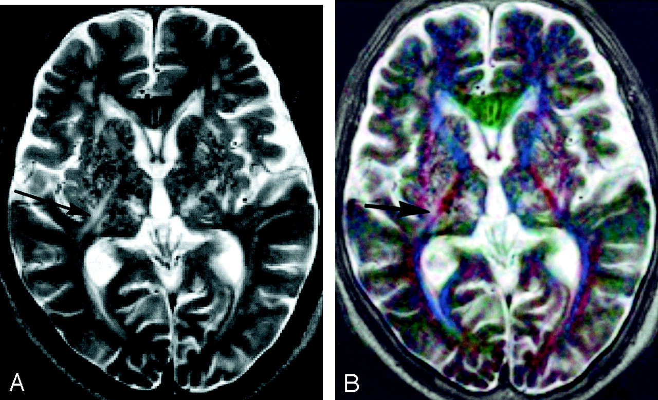
  • Fig 2.
    • Download figure
    • Open in new tab
    • Download powerpoint
    Fig 2.

    Images of a 63-year-old man with right parietal glioblastoma.

    A, Conventional fast spin-echo T2-weighted image shows peritumoral white matter edema around the right lateral ventricle that is pressed down by the tumor. The precise location of the right corona radiata and the posterior limb of the left internal capsule are not apparent on this image.

    B, Color-coded image constructed from three inverted orthogonal diffusion-weighted images. Fibers running superoinferiorly, anteroposteriorly, and transversely are displayed with red, blue, and green, respectively. Because the T2 shine-through phenomenon counteracts the contrast caused by diffusion, the peritumoral edema is not distinct from normal brain.

    C, Color-coded image created from three orthogonal ADC maps.

    D, Image after registration and addition of the images shown in A and B has a contrast similar to and a quality superior to those of the image shown in C. The right deep white matter with edema is clearly resolved into three parts: from medial to lateral, the corpus callosum (green, arrow), corona radiata (orange, arrowhead), and superior longitudinal fasciculus (blue, open arrow). The posterior limb of the left internal capsule can be easily identified by the red hue (open arrowhead).

  • Fig 3.
    • Download figure
    • Open in new tab
    • Download powerpoint
    Fig 3.

    Superposed color images obtained in a 41-year-old woman with dyskinesia. Diagnosis has not yet been established.

    A, Axial image. The white matter lateral to the posterior horn of the lateral ventricle appears to be divided into four layers. They are probably, from medial to lateral, the tapetum (red, arrowheads), internal sagittal stratum (dark blue, open arrowheads), external sagittal stratum (bright blue, arrows), and white matter containing the superior longitudinal fasciculus (red, open arrows). The pulvinar of the thalamus appears yellowish green (*).

    B, Coronal image. The posterior limbs of the internal capsules are identified as red bands (arrowheads). The external and the extreme capsules (cannot be separated) are seen as reddish sheets (arrows). The superior longitudinal fasciculi appear greenish blue (open arrows). The cingula appear blue (open arrowheads). The inferolateral parts of the temporal lobes show artifact because of the failure to correct severe distortion.
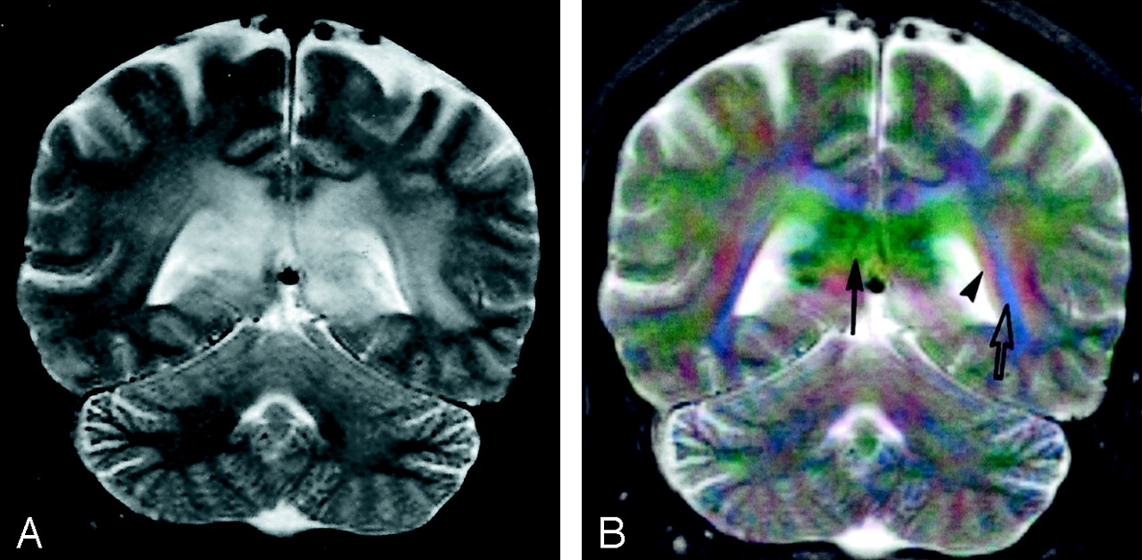
  • Fig 4.
    • Download figure
    • Open in new tab
    • Download powerpoint
    Fig 4.

    Images obtained in an 84-year-old man with multiple lacunar hemorrhages and infarcts.

    A, Spin-echo T2-weighted image shows multiple small foci of hyper- and hypointensity in the basal ganglia and thalami. Linear hyperintensity (arrow) is seen near the right posterior limb of the internal capsule. Spatial relations between the internal capsule and these lesions are unclear.

    B, On the superposed image, the posterior limbs and anterior limbs of the internal capsule appear red and blue, respectively. They are easy to identify, and the right internal capsule is situated medially to the linear hyperintensity (arrow).

  • Fig 5.
    • Download figure
    • Open in new tab
    • Download powerpoint
    Fig 5.

    Images of a 6-month-old male patient with epilepsy. No abnormality is found by MR imaging examination.

    A, Spin-echo T2-weighted image.

    B, Superposed color image. The white matter anterolateral to the inferior horn of the lateral ventricle appears red (arrows). This is probably part of the uncinate fasciculus running up from the temporal pole to the inferior margin of the extreme and external capsule. The pyramidal tract in the basis pontis (red, open arrows) is distinguished from the transverse pontine (pontocerebellar) fiber (green) by the difference in hue. Both the inferior (lateral part) and superior (medial part) cerebellar peduncles appear red (arrowheads) between the fourth ventricle and the middle cerebellar peduncle (blue, open arrowheads).

  • Fig 6.
    • Download figure
    • Open in new tab
    • Download powerpoint
    Fig 6.

    Coronal view images of a 45-year-old woman with lymphoma in the splenium of the corpus callosum.

    A, Spin-echo T2-weighted image shows white matter edema around the lateral ventricles. The corpus callosum cannot be clearly recognized.

    B, Superposed color image. The corpus callosum is clearly identified as a green structure (arrow). The white matter lateral to the left lateral ventricle appears to have at least three layers: medial to lateral, red (tapetum, arrowhead), blue (internal and external sagittal strata, open arrow), and red layers. The asymmetrically widened tapetum on the left indicates extension of peritumoral edema in this region.

  • Fig 7.
    • Download figure
    • Open in new tab
    • Download powerpoint
    Fig 7.

    Left sphenoid ridge meningioma in a 54-year-old woman.

    A, Spin-echo T2-weighted image.

    B, Superposed color image shows the meningioma with certain colors: the anterior and posterior parts appear violet, and the left and right parts are green, which can be interpreted (although it has not been confirmed histologically) to mean that this tumor, as a whole, has a radially oriented internal structure. The posterior limb of the left internal capsule (arrow) appears redder and thinner than the contralateral limb (arrowhead). This indicates that the tumor compresses the limb and deflects its direction vertically.
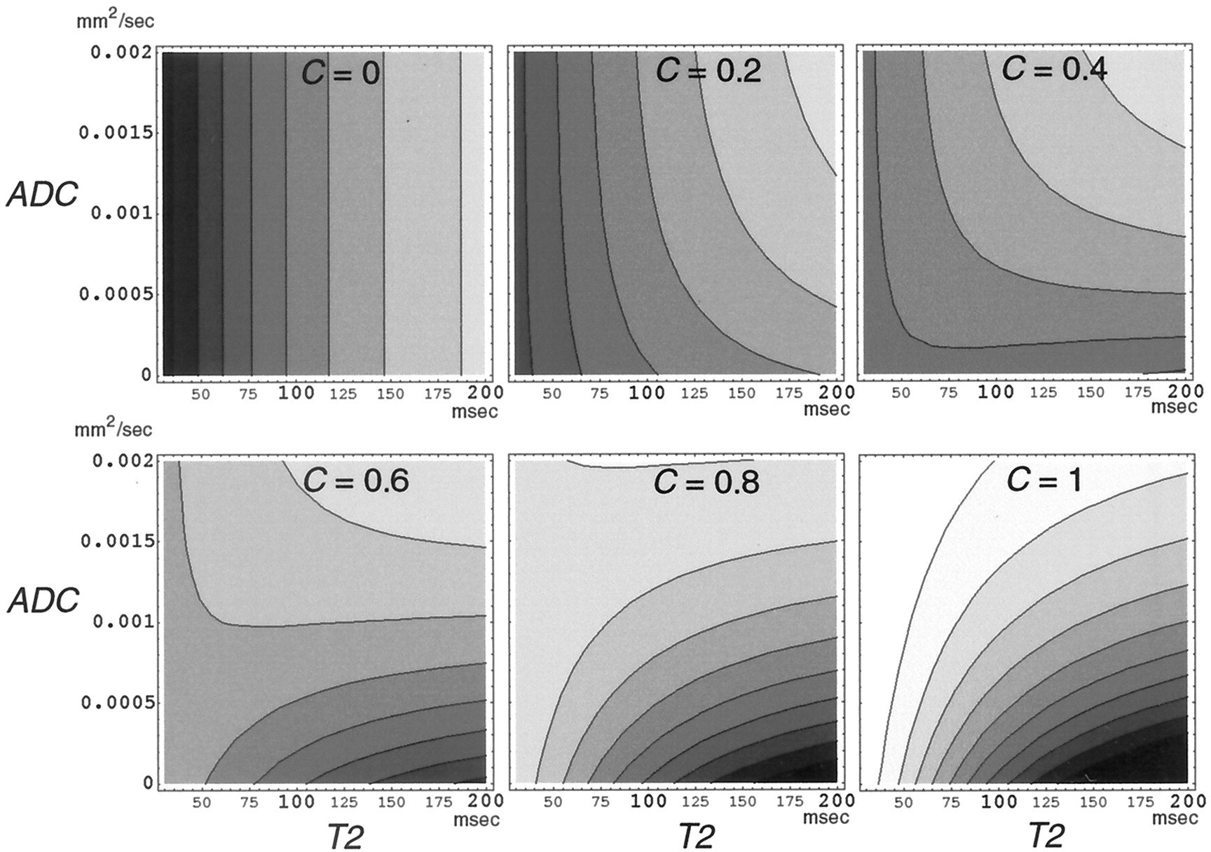
  • Fig 8.
    • Download figure
    • Open in new tab
    • Download powerpoint
    Fig 8.

    Brightness of a color channel of the fused image (fusion of spin-echo T2-weighted image and inverted diffusion-weighted image) calculated from equation 1. The brightness is plotted with gray levels for T2 (x axis) versus ADC (y axis) for the weighting factor, C = 0, 0.2, 0.4, 0.6, 0.8, and 1. The brightness for C = 0 or C = 1 contains only the spin-echo T2-weighted contrast or the inverted diffusion-weighted contrast, respectively.

PreviousNext
Back to top

In this issue

American Journal of Neuroradiology: 24 (3)
American Journal of Neuroradiology
Vol. 24, Issue 3
1 Mar 2003
  • Table of Contents
  • Index by author
Advertisement
Print
Download PDF
Email Article

Thank you for your interest in spreading the word on American Journal of Neuroradiology.

NOTE: We only request your email address so that the person you are recommending the page to knows that you wanted them to see it, and that it is not junk mail. We do not capture any email address.

Enter multiple addresses on separate lines or separate them with commas.
Practical Visualization of Internal Structure of White Matter for Image Interpretation: Staining a Spin-Echo T2-Weighted Image with Three Echo-Planar Diffusion-Weighted Images
(Your Name) has sent you a message from American Journal of Neuroradiology
(Your Name) thought you would like to see the American Journal of Neuroradiology web site.
CAPTCHA
This question is for testing whether or not you are a human visitor and to prevent automated spam submissions.
Cite this article
Hajime Tamura, Shoki Takahashi, Noriko Kurihara, Shogo Yamada, Jun Hatazawa, Toshio Okudera
Practical Visualization of Internal Structure of White Matter for Image Interpretation: Staining a Spin-Echo T2-Weighted Image with Three Echo-Planar Diffusion-Weighted Images
American Journal of Neuroradiology Mar 2003, 24 (3) 401-409;

Citation Manager Formats

  • BibTeX
  • Bookends
  • EasyBib
  • EndNote (tagged)
  • EndNote 8 (xml)
  • Medlars
  • Mendeley
  • Papers
  • RefWorks Tagged
  • Ref Manager
  • RIS
  • Zotero
0 Responses
Respond to this article
Share
Bookmark this article
Practical Visualization of Internal Structure of White Matter for Image Interpretation: Staining a Spin-Echo T2-Weighted Image with Three Echo-Planar Diffusion-Weighted Images
Hajime Tamura, Shoki Takahashi, Noriko Kurihara, Shogo Yamada, Jun Hatazawa, Toshio Okudera
American Journal of Neuroradiology Mar 2003, 24 (3) 401-409;
del.icio.us logo Twitter logo Facebook logo Mendeley logo
  • Tweet Widget
  • Facebook Like
  • Google Plus One

Jump to section

  • Article
    • Abstract
    • Methods
    • Results
    • Discussion
    • Conclusion
    • Appendix A
    • Appendix B
    • References
  • Figures & Data
  • Info & Metrics
  • Responses
  • References
  • PDF

Related Articles

  • No related articles found.
  • PubMed
  • Google Scholar

Cited By...

  • No citing articles found.
  • Crossref
  • Google Scholar

This article has not yet been cited by articles in journals that are participating in Crossref Cited-by Linking.

More in this TOC Section

  • Usefulness of Quantitative Susceptibility Mapping for the Diagnosis of Parkinson Disease
  • White Matter Alterations in the Brains of Patients with Active, Remitted, and Cured Cushing Syndrome: A DTI Study
  • Qualitative and Quantitative Analysis of MR Imaging Findings in Patients with Middle Cerebral Artery Stroke Implanted with Mesenchymal Stem Cells
Show more BRAIN

Similar Articles

Advertisement

Indexed Content

  • Current Issue
  • Accepted Manuscripts
  • Article Preview
  • Past Issues
  • Editorials
  • Editor's Choice
  • Fellows' Journal Club
  • Letters to the Editor
  • Video Articles

Cases

  • Case Collection
  • Archive - Case of the Week
  • Archive - Case of the Month
  • Archive - Classic Case

Special Collections

  • AJNR Awards
  • ASNR Foundation Special Collection
  • Most Impactful AJNR Articles
  • Photon-Counting CT
  • Spinal CSF Leak Articles (Jan 2020-June 2024)

More from AJNR

  • Trainee Corner
  • Imaging Protocols
  • MRI Safety Corner

Multimedia

  • AJNR Podcasts
  • AJNR Scantastics

Resources

  • Turnaround Time
  • Submit a Manuscript
  • Submit a Video Article
  • Submit an eLetter to the Editor/Response
  • Manuscript Submission Guidelines
  • Statistical Tips
  • Fast Publishing of Accepted Manuscripts
  • Graphical Abstract Preparation
  • Imaging Protocol Submission
  • Evidence-Based Medicine Level Guide
  • Publishing Checklists
  • Author Policies
  • Become a Reviewer/Academy of Reviewers
  • News and Updates

About Us

  • About AJNR
  • Editorial Board
  • Editorial Board Alumni
  • Alerts
  • Permissions
  • Not an AJNR Subscriber? Join Now
  • Advertise with Us
  • Librarian Resources
  • Feedback
  • Terms and Conditions
  • AJNR Editorial Board Alumni

American Society of Neuroradiology

  • Not an ASNR Member? Join Now

© 2025 by the American Society of Neuroradiology All rights, including for text and data mining, AI training, and similar technologies, are reserved.
Print ISSN: 0195-6108 Online ISSN: 1936-959X

Powered by HighWire