
Article Figures & Data
Figures
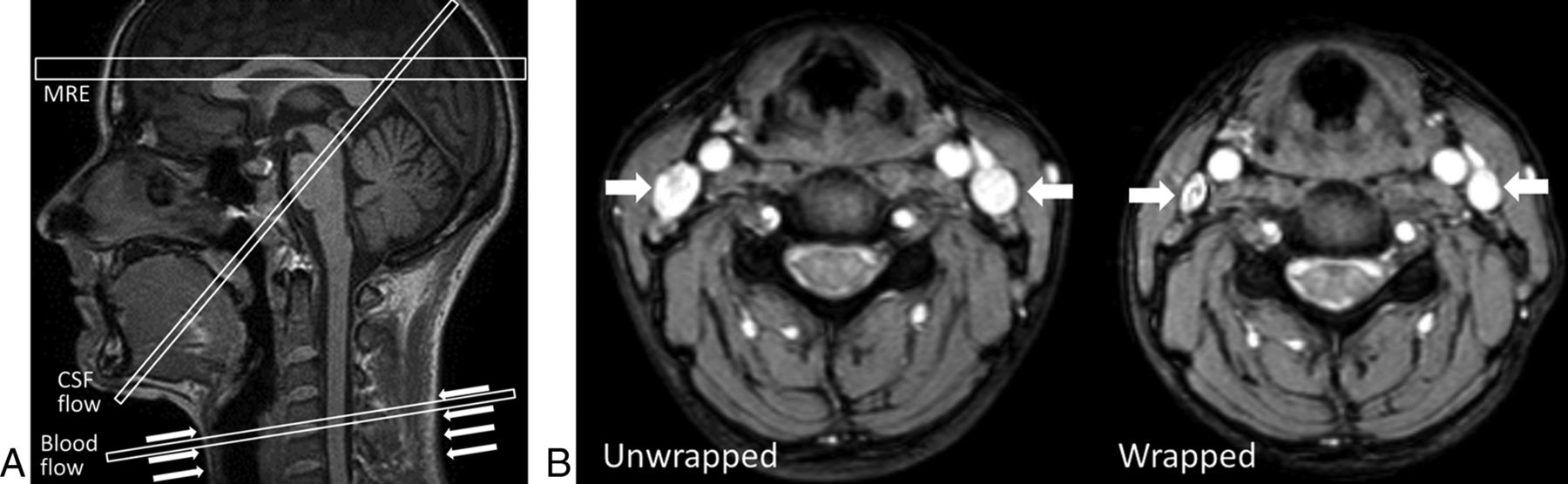
- Fig 1.
A, Sagittal view of imaging planes for the MRE study within and just above the ventricles, phase-contrast MR imaging CSF flow study of the cerebral aqueduct, and phase-contrast MR imaging flow study of the blood flow in the neck vessels at the level of the compression. The arrows denote the position of neck wrap. B, Images of neck vessels with and without the neck wrap, showing the reduction in internal jugular vein area. Internal jugular veins are indicated by white arrows.
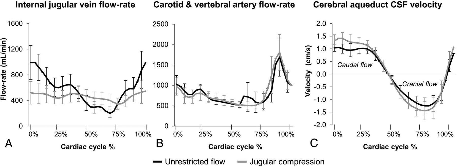
- Fig 2.
Group mean flow waveforms for internal jugular vein flow (A) and carotid and vertebral artery flow (B); and group mean CSF velocity waveforms through the cerebral aqueduct (C). Venous flow-rate peaks are suppressed with jugular compression; however, arterial flow waveforms are unaffected. Maximal caudally directed velocity of the CSF in the aqueduct increases with jugular compression.
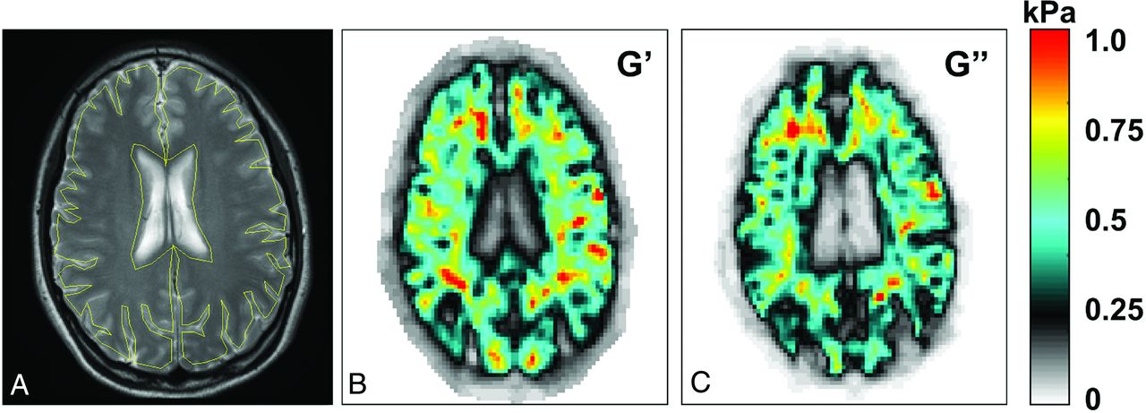
- Fig 3.
ROI and sample viscoelastic maps. A, Sample high-resolution anatomic image shows the ROI (yellow line). Viscoelastic property maps for G′ (B) and G″ (C) in kilopascals. In the viscoelastic maps, the ventricles and large sulci have very low (near zero) shear moduli, indicating that they are filled with CSF, which has waterlike properties. Stiffer tissue is indicated by warmer colors, as indicated by the color bar (right).
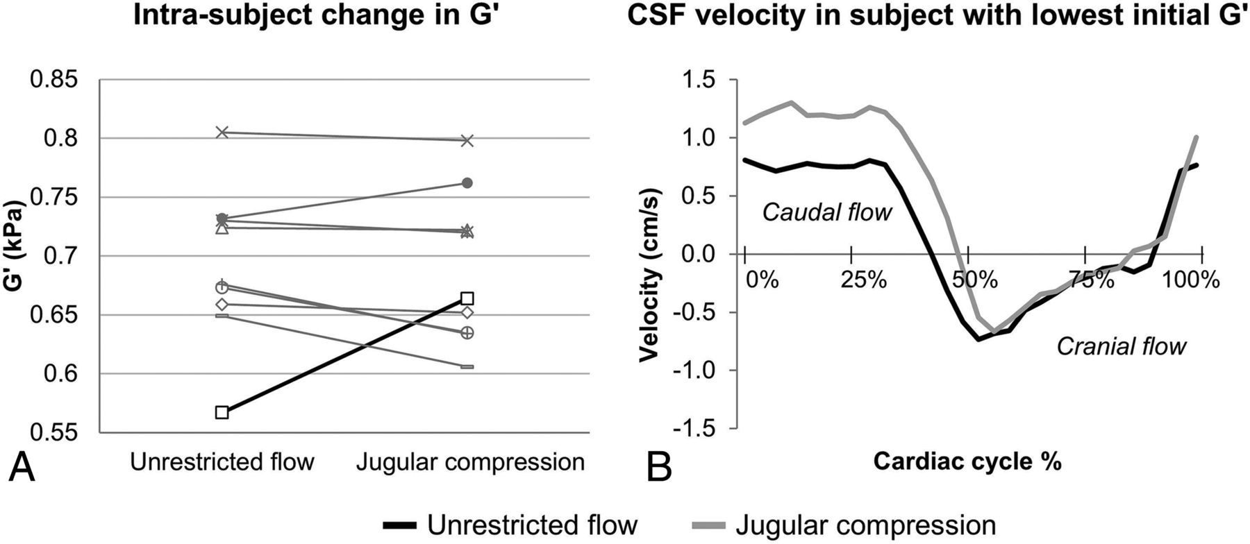
- Fig 4.
A, Individual subject change in G′ in unrestricted and jugular compression conditions for all subjects. B, Cerebral aqueduct CSF velocity waveforms for a single subject are depicted by the black line in A. In this subject, the 17% increase in G′ with jugular compression was related to a 61% increase in the maximal caudal CSF velocity and an 11% longer caudal-flow duration.
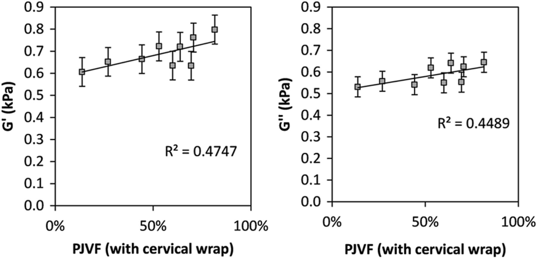
- Fig 5.
The relationship between shear moduli and the percentage of jugular vein flow with the cervical wrap in place. Storage modulus (G′, left) and loss modulus (G″, right). Error bars are standard error of the mean in both panels. Both storage and loss moduli are significantly correlated with the percentage of jugular vein flow with the cervical wrap in place (P < .05).


Tables
Internal Jugular Vein Flow Arterial Flow Unrestricted Flow Jugular Compression Unrestricted Flow Jugular Compression Mean vessel area (cm2) Mean 1.17 ± 0.54 0.70 ± 0.35 1.0 ± 0.77 0.97 ± 0.68 Range 0.40–2.18 0.29–1.41 −0.99–1.53 −0.76–1.56 t test t = 3.56, P = .005a t = 0.51, P = .604 Mean flow rate (mL/min) (caudal flow for venous flow, cranial for arterial) Mean 572.2 ± 219.7 460.8 ± 202.4 814.7 ± 221.9 815.3 ± 290.6 Range 153.8–862.2 135–726.4 268–989.2 127.7–1070 t test t = 2.85, P = .008a −0.01, P = .989 Flow-rate amplitude (mL/min) Mean 974.1 ± 523.6 382.6 ± 312.7 1803.8 ± 490.1 1720.6 ± 688.4 Range 198.5–1838.8 56.8–1069.0 601.4–2290.2 282.8–2721.4 t test t = 3.75, P = .002a t = 0.58, P = .558 ↵a P < .05, paired t test.
PJVF Unrestricted Flow Jugular Compression Subject 1 97.2% 53.0% Subject 2 84.1% 70.6% Subject 3 82.1% 81.5% Subject 4 76.8% 69.5% Subject 5 74.2% 44.3% Subject 6 61.2% 27.0% Subject 7 59.8% 63.8% Subject 8 59.3% 60.0% Subject 9 17.9% 13.7% Mean 68.1 ± 22.7% 53.7 ± 21.9% t test t = 2.33, P = .019a Predominantly jugular flow (between 50% and 100%) Subjects 8/9 6/9 Predominantly nonjugular flow (<50%) Subjects 1/9 3/9 ↵a P < .05, paired t test.
Aqueductal Flow Unrestricted Flow Jugular Compression Maximum caudal velocity (cm/s) Mean 1.18 ± 0.33 1.53 ± 0.42 Range 0.81–1.85 1.04–2.36 t test t = −4.96, P = .0004a Maximum cranial velocity (cm/s) Mean 1.38 ± 0.57 1.62 ± 0.62 Range 0.73–2.47 0.66–2.48 t test t = 1.58, P = .066 Duration of caudally directed flow (% cardiac cycle) Mean 49.9 ± 5.2% 51.4 ± 5.3% Range 40.7%–57.7% 42.1%–61.7% t test t = −1.0, P = .160 ↵a P < .05, paired t test.
Unrestricted Flow Jugular Compression G′ (kPa) Mean 0.691 ± 0.067 0.688 ± 0.065 Range 0.567–0.805 0.606–0.798 t test t = 0.17, P = .436 G″ (kPa) Mean 0.587 ± 0.052 0.585 ± 0.046 Range 0.482–0.662 0.531–0.645 t test t = 0.23, P = .413 - Table 5:
Linear regression coefficients for the relationships between PJVF and G′ and G″ with unrestricted flow and with jugular compression, and change in mean G′, G″, and maximal caudal CSF velocity
R2 P G′ and PJVF, unrestricted flow 0.136 .330 G′ and PJVF, jugular compression 0.475 .040a G″ and PJVF, unrestricted flow 0.151 .301 G″ and PJVF, jugular compression 0.449 .048a % Change G′ and % change in max. caudal CSF velocity 0.412 .063 % Change G″ and % change in max. caudal CSF velocity 0.171 .268 Note:—max. indicates maximum.
↵a P < .05, paired t test.


Jump to section
Related Articles
Cited By...
- Differences in the Calculated Transvenous Pressure Drop between Chronic Hydrocephalus and Idiopathic Intracranial Hypertension
- Analysis of head impact exposure and brain microstructure response in a season-long application of a jugular vein compression collar: a prospective, neuroimaging investigation in American football