
Article Figures & Data
Figures
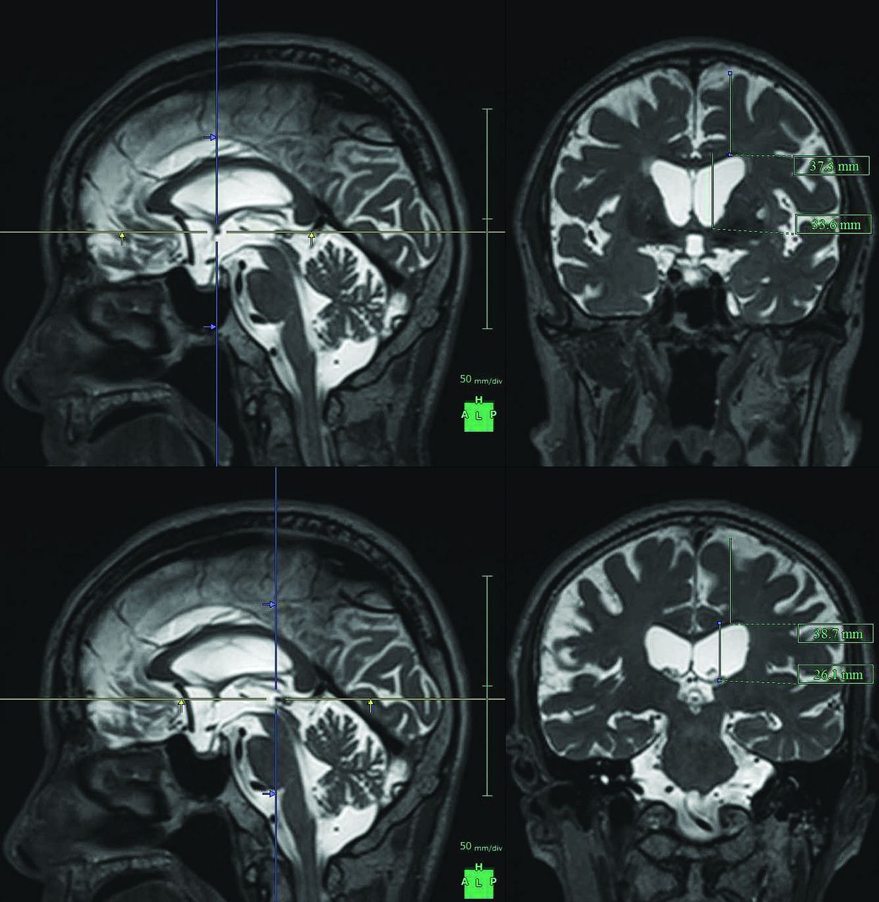
- Fig 1.
Maximum widths of the lateral ventricles and brain substances just above the lateral ventricles. The figures show the multiplanar reconstruction images on the T2-weighted 3D SPACE sequence. The crosses of the blue and yellow lines on the left figures indicate the points of the anterior commissure (upper) and posterior commissure (lower), and the right figures show the coronal planes at the blue lines of the left figures.
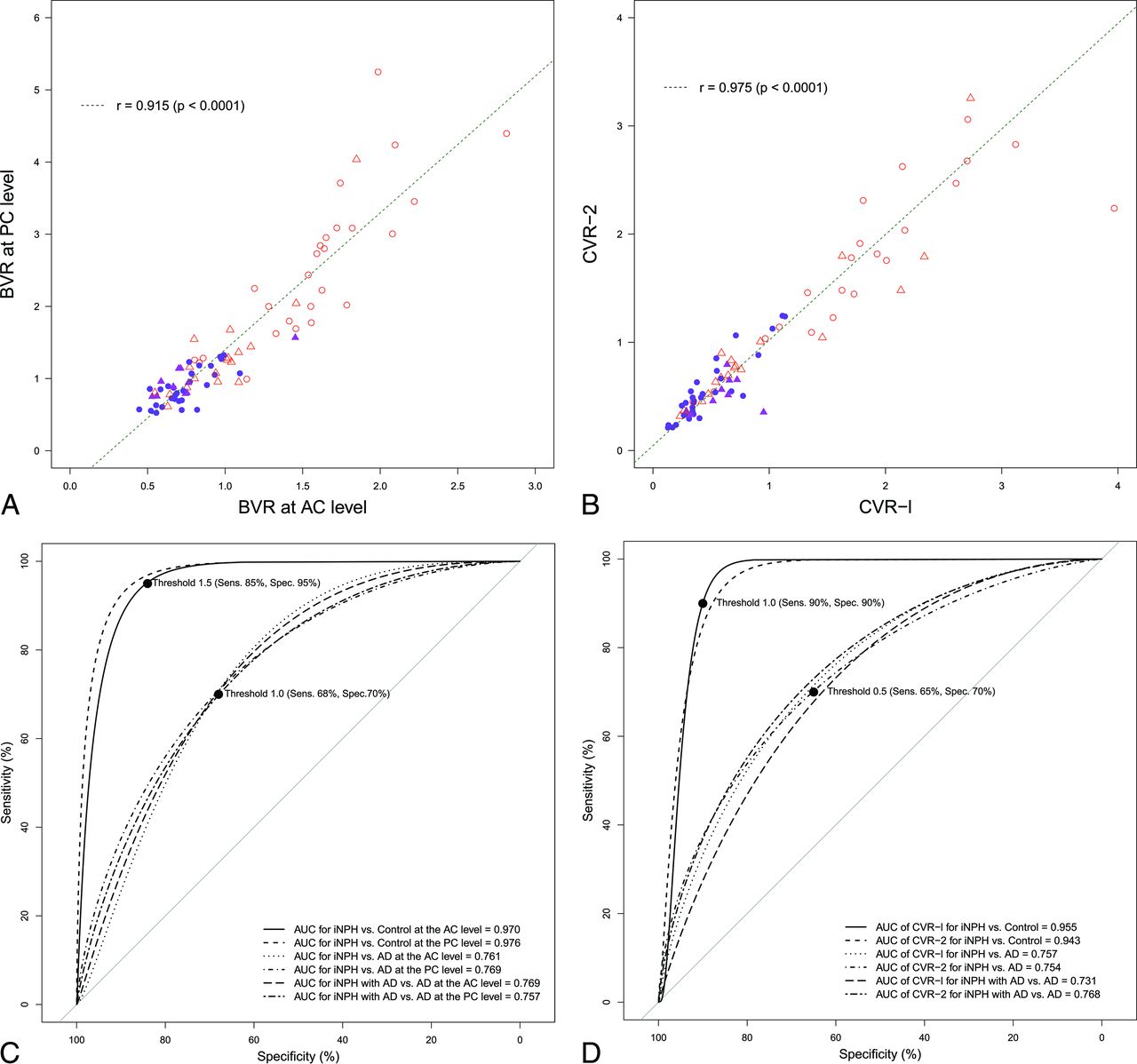
- Fig 2.
Scatterplots and receiver operating characteristic curves for the differential diagnosis of idiopathic normal pressure hydrocephalus and Alzheimer disease. The left upper diagram (A) shows the scatterplot and linear regression for the correlation between the BVR at the anterior level (x-axis) and those at the posterior commissure level (y-axis). The right upper diagram (B) shows the scatterplot of the 3D volumetric convexity subarachnoid space to ventricle ratio-1 (CVR-1) and CVR-2. The purple circle indicates iNPH, the purple triangle indicates iNPH concurrent with Alzheimer disease, the open red triangle indicates AD, and the open red circle indicates control. The lower diagram shows the receiver operating characteristic graph curves of BVR (left, C), CVR-1, and CVR-2 (right, D) for the differential diagnosis of iNPH or iNPH with AD from AD or controls. The x-axis shows specificity, and the y-axis shows sensitivity. The black marks indicate the point of the maximum area under the receiver operating characteristic curve and optimal thresholds (sensitivity and specificity). The maximum areas under the receiver operating characteristic curves for each comparison are displayed in the lower graph.
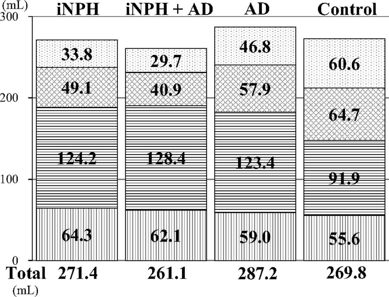
- Fig 3.
Mean volumes of the total ventricle and total subarachnoid space. The mean volumes of the total ventricle are displayed in a downward direction (dotted pattern). The mean volumes of the total subarachnoid spaces are displayed after division of the upper (vertical striped pattern) and lower parts (wave pattern) in a horizontal section on the anterior/posterior commissure plane at the level of the junction point of the vein of Galen and straight sinus.
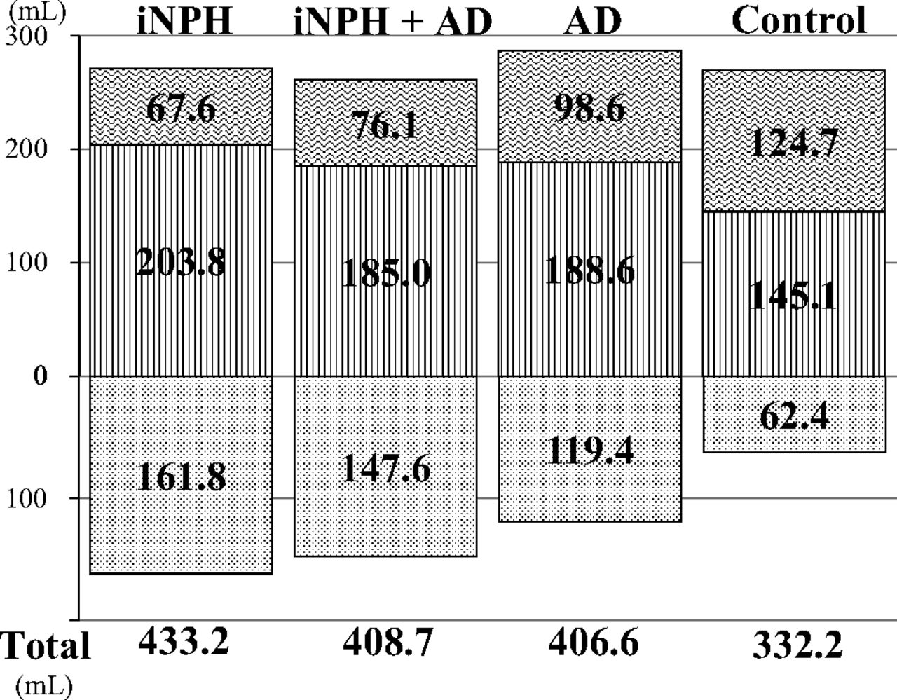
- Fig 4.
The mean volumes of the segmented parts of the subarachnoid spaces. The vertical-striped pattern indicates the subarachnoid space in the posterior fossa. The horizontal-striped pattern indicates the subarachnoid space in the basal cistern and Sylvian fissure, the checked pattern indicates the subarachnoid space in the frontal convexity subarachnoid space, and the dotted pattern indicates the subarachnoid space in the parietal convexity subarachnoid space.

Tables
iNPH (n = 30) iNPH + AD (n = 10) AD (n = 18) Controls (n = 26) P1a P2b Width of the ventricle at AC level (mm) 41.2 ± 5.0 41.0 ± 5.9 35.6 ± 5.8 27.2 ± 6.2 .002 .014 Width of the brain at AC level (mm) 29.7 ± 3.5 29.3 ± 3.6 33.2 ± 4.6 40.7 ± 4.2 .006 .029 Width of the ventricle at PC level (mm) 35.3 ± 6.1 32.9 ± 4.7 28.7 ± 6.9 18.8 ± 6.2 .002 .027 Width of the brain at PC level (mm) 29.6 ± 3.9 31.0 ± 3.0 34.0 ± 5.3 41.9 ± 4.7 .010 .150 BVR at AC level 0.74 ± 0.16 0.75 ± 0.26 0.98 ± 0.31 1.59 ± 0.45 <.001 <.001 BVR at PC level 0.88 ± 0.25 0.97 ± 0.25 1.34 ± 0.76 2.54 ± 1.08 <.001 <.001 Evans index 0.34 ± 0.04 0.34 ± 0.37 0.32 ± 0.40 0.28 ± 3.8 .079 .286 z-Evans index 0.43 ± 0.04 0.43 ± 0.61 0.38 ± 0.61 0.29 ± 6.3 .001 .016 Callosal angle (degree) 65.0 ± 20.2 61.0 ± 15.5 86.1 ± 24.0 103.7 ± 15.9 .005 .010 Total intracranial volume (mL) 1519 ± 127 1457 ± 139 1513 ± 200 1484 ± 146 .741 .443 Total CSF volume (mL) 433.2 ± 82.0 408.7 ± 105 406.6 ± 97.1 332.2 ± 112 .428 .654 Brain parenchyma volume (mL) 1086 ± 85.4 1048 ± 120 1106 ± 145 1152 ± 157 .358 .175


Jump to section
Related Articles
Cited By...
- CSF circulation and dispersion yield rapid clearance from intracranial compartments
- Variability of Normal Pressure Hydrocephalus Imaging Biomarkers with Respect to Section Plane Angulation: How Wrong a Radiologist Can Be?
- Quantification of Oscillatory Shear Stress from Reciprocating CSF Motion on 4D Flow Imaging