Advertisement

Main menu

  • Home
  • Content
    • Current Issue
    • Accepted Manuscripts
    • Article Preview
    • Past Issue Archive
    • Video Articles
    • AJNR Case Collection
    • Case of the Week Archive
    • Case of the Month Archive
    • Classic Case Archive
  • Special Collections
    • AJNR Awards
    • ASNR Foundation Special Collection
    • Most Impactful AJNR Articles
    • Photon-Counting CT
    • Spinal CSF Leak Articles (Jan 2020-June 2024)
  • Multimedia
    • AJNR Podcasts
    • AJNR SCANtastic
    • Trainee Corner
    • MRI Safety Corner
    • Imaging Protocols
  • For Authors
    • Submit a Manuscript
    • Submit a Video Article
    • Submit an eLetter to the Editor/Response
    • Manuscript Submission Guidelines
    • Statistical Tips
    • Fast Publishing of Accepted Manuscripts
    • Graphical Abstract Preparation
    • Imaging Protocol Submission
    • Author Policies
  • About Us
    • About AJNR
    • Editorial Board
    • Editorial Board Alumni
  • More
    • Become a Reviewer/Academy of Reviewers
    • Subscribers
    • Permissions
    • Alerts
    • Feedback
    • Advertisers
    • ASNR Home

User menu

  • Alerts
  • Log in

Search

  • Advanced search
American Journal of Neuroradiology

American Journal of Neuroradiology

ASHNR American Society of Functional Neuroradiology ASHNR American Society of Pediatric Neuroradiology ASSR
  • Alerts
  • Log in

Advanced Search

  • Home
  • Content
    • Current Issue
    • Accepted Manuscripts
    • Article Preview
    • Past Issue Archive
    • Video Articles
    • AJNR Case Collection
    • Case of the Week Archive
    • Case of the Month Archive
    • Classic Case Archive
  • Special Collections
    • AJNR Awards
    • ASNR Foundation Special Collection
    • Most Impactful AJNR Articles
    • Photon-Counting CT
    • Spinal CSF Leak Articles (Jan 2020-June 2024)
  • Multimedia
    • AJNR Podcasts
    • AJNR SCANtastic
    • Trainee Corner
    • MRI Safety Corner
    • Imaging Protocols
  • For Authors
    • Submit a Manuscript
    • Submit a Video Article
    • Submit an eLetter to the Editor/Response
    • Manuscript Submission Guidelines
    • Statistical Tips
    • Fast Publishing of Accepted Manuscripts
    • Graphical Abstract Preparation
    • Imaging Protocol Submission
    • Author Policies
  • About Us
    • About AJNR
    • Editorial Board
    • Editorial Board Alumni
  • More
    • Become a Reviewer/Academy of Reviewers
    • Subscribers
    • Permissions
    • Alerts
    • Feedback
    • Advertisers
    • ASNR Home
  • Follow AJNR on Twitter
  • Visit AJNR on Facebook
  • Follow AJNR on Instagram
  • Join AJNR on LinkedIn
  • RSS Feeds

AJNR is seeking candidates for the AJNR Podcast Editor. Read the position description.

Research ArticleADULT BRAIN
Open Access

Quantitative Susceptibility Mapping and R2* Measured Changes during White Matter Lesion Development in Multiple Sclerosis: Myelin Breakdown, Myelin Debris Degradation and Removal, and Iron Accumulation

Y. Zhang, S.A. Gauthier, A. Gupta, W. Chen, J. Comunale, G.C.-Y. Chiang, D. Zhou, G. Askin, W. Zhu, D. Pitt and Y. Wang
American Journal of Neuroradiology September 2016, 37 (9) 1629-1635; DOI: https://doi.org/10.3174/ajnr.A4825
Y. Zhang
aFrom the Department of Radiology (Y.Z., W.C., W.Z.), Tongji Hospital, Tongji Medical College, Huazhong University of Science and Technology, Wuhan, China
bDepartments of Radiology (Y.Z., A.G., J.C., G.C.-Y.C., D.Z., Y.W.)
  • Find this author on Google Scholar
  • Find this author on PubMed
  • Search for this author on this site
  • ORCID record for Y. Zhang
S.A. Gauthier
cNeurology (S.A.G.)
  • Find this author on Google Scholar
  • Find this author on PubMed
  • Search for this author on this site
  • ORCID record for S.A. Gauthier
A. Gupta
bDepartments of Radiology (Y.Z., A.G., J.C., G.C.-Y.C., D.Z., Y.W.)
  • Find this author on Google Scholar
  • Find this author on PubMed
  • Search for this author on this site
  • ORCID record for A. Gupta
W. Chen
aFrom the Department of Radiology (Y.Z., W.C., W.Z.), Tongji Hospital, Tongji Medical College, Huazhong University of Science and Technology, Wuhan, China
  • Find this author on Google Scholar
  • Find this author on PubMed
  • Search for this author on this site
  • ORCID record for W. Chen
J. Comunale
bDepartments of Radiology (Y.Z., A.G., J.C., G.C.-Y.C., D.Z., Y.W.)
  • Find this author on Google Scholar
  • Find this author on PubMed
  • Search for this author on this site
  • ORCID record for J. Comunale
G.C.-Y. Chiang
bDepartments of Radiology (Y.Z., A.G., J.C., G.C.-Y.C., D.Z., Y.W.)
  • Find this author on Google Scholar
  • Find this author on PubMed
  • Search for this author on this site
  • ORCID record for G.C.-Y. Chiang
D. Zhou
bDepartments of Radiology (Y.Z., A.G., J.C., G.C.-Y.C., D.Z., Y.W.)
  • Find this author on Google Scholar
  • Find this author on PubMed
  • Search for this author on this site
  • ORCID record for D. Zhou
G. Askin
dHealthcare Policy and Research (G.A.), Weill Cornell Medical College, New York, New York
  • Find this author on Google Scholar
  • Find this author on PubMed
  • Search for this author on this site
  • ORCID record for G. Askin
W. Zhu
aFrom the Department of Radiology (Y.Z., W.C., W.Z.), Tongji Hospital, Tongji Medical College, Huazhong University of Science and Technology, Wuhan, China
  • Find this author on Google Scholar
  • Find this author on PubMed
  • Search for this author on this site
  • ORCID record for W. Zhu
D. Pitt
eDepartment of Neurology (D.P.), School of Medicine, Yale University, New Haven, Connecticut
  • Find this author on Google Scholar
  • Find this author on PubMed
  • Search for this author on this site
  • ORCID record for D. Pitt
Y. Wang
bDepartments of Radiology (Y.Z., A.G., J.C., G.C.-Y.C., D.Z., Y.W.)
fDepartment of Biomedical Engineering (Y.W.), Cornell University, Ithaca, New York.
  • Find this author on Google Scholar
  • Find this author on PubMed
  • Search for this author on this site
  • ORCID record for Y. Wang
  • Article
  • Figures & Data
  • Info & Metrics
  • Responses
  • References
  • PDF
Loading

Article Figures & Data

Figures

  • Tables
  • Fig 1.
    • Download figure
    • Open in new tab
    • Download powerpoint
    Fig 1.

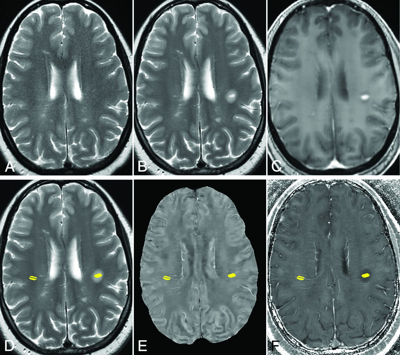
    Example of ROIs of an MS lesion and reference at normal-appearing WM in a 44-year-old woman with MS. A, T2WI 8 months before the appearance of the enhancing lesion. B, T2WI, and C, T1WI + Gd image of 1 enhancing MS lesion. D, T2WI, E, QSM, and F, R2* images with ROIs of the enhancing lesion (left side) and the normal-appearing WM (right side). The vein inside the selected normal-appearing WM is excluded.

  • Fig 2.
    • Download figure
    • Open in new tab
    • Download powerpoint
    Fig 2.

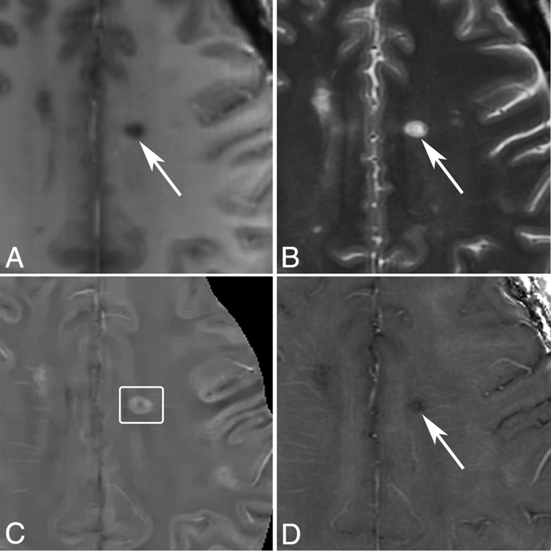
    MR images of a nodular-enhancing lesion in a 43-year-old man with MS. A, T1WI + Gd. B, T2WI. C, QSM. D, R2*. A nodular-enhancing lesion (arrows) is found on the T1WI + Gd and appears QSM isointense (C, box).

  • Fig 3.
    • Download figure
    • Open in new tab
    • Download powerpoint
    Fig 3.

    MR images of a shell-enhancing lesion in a 51-year-old man with MS. A, T1WI + Gd. B, T2WI. C, QSM. D, R2*. A shell-enhancing lesion (arrows) is found on the T1WI + Gd and appears slightly QSM hyperintense (C, box).

  • Fig 4.
    • Download figure
    • Open in new tab
    • Download powerpoint
    Fig 4.

    MR images of new T2 nonenhancing lesions (< 0.7 years old) in a 42-year-old man with MS. A, T1WI + Gd. B, T2WI. C, QSM. D, R2*. More than 3 new nonenhancing lesions (arrows) are found by comparing with the former MR imaging 0.7 years ago. All of them appear QSM hyperintense (C, box) and have hyperintense rims on R2*.

  • Fig 5.
    • Download figure
    • Open in new tab
    • Download powerpoint
    Fig 5.

    MR images of a nonenhancing lesion that is 1.2 years old in a 48-year-old woman with MS. A, T1WI + Gd. B, T2WI. C, QSM. D, R2*. One lesion appears hyperintense with a thick rim on QSM and hypointense on R2*.

  • Fig 6.
    • Download figure
    • Open in new tab
    • Download powerpoint
    Fig 6.

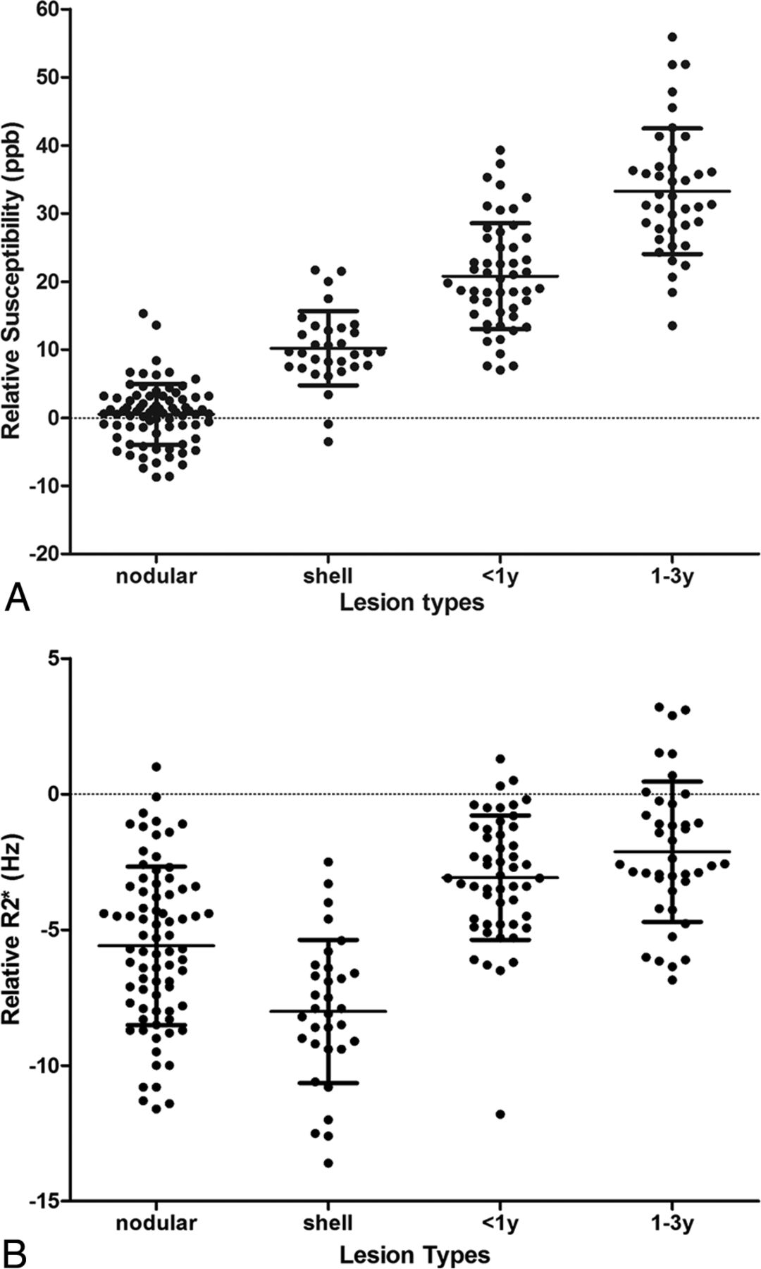
    Scatterplot of susceptibility relative to normal-appearing WM measured on A, QSM and B, R2*. ppb indicates parts per billion.

Tables

  • Figures
    • View popup
    Table 1:

    Quantitative susceptibility and R2* values for nodular-enhancing, shell-enhancing, nonenhancing < 1 year, and nonenhancing 1–3 years old lesions

    NodularShellNonenhancing < 1 YearNonenhancing 1–3 Years
    No. of lesions77324841
    No. of patients32212720
    Susceptibility relative to NAWM, ppb0.5 ± 4.410.2 ± 5.420.2 ± 7.833.2 ± 8.2
    R2* values relative to NAWM, Hz−5.6 ± 2.9−8.0 ± 2.6−3.1 ± 2.3−2.0 ± 2.6
    • Note:—NAWM indicates normal-appearing white matter; ppb, parts per billion.

    • View popup
    Table 2:

    Statistical comparison of relative susceptibility from 4 groups of lesionsa

    ComparisonNodularShell<1 year
    Shell−9.8621, (−11.7759, −7.9484), P < .0001––
    <1 year−20.3123, (−23.0288, −17.5959), P < .0001−10.4502, (−13.6101, −7.2903), P < .0001–
    1–3 years−32.6287, (−35.9416, −29.3159), P < .0001−22.7666, (−26.2751, −19.2581), P < .0001−12.3164, (−16.3345, −8.2983), P < .0001
    • ↵a Data given as relative parameter estimates (β), (95% confidence limits), P value. Parameter estimates, 95% CI, and SE from multivariable generalized estimating equation, controlling for age, Expanded Disability Status Scale score, and disease duration. β estimates were obtained by setting the reference category to different lesion group.

    • View popup
    Table 3:

    Statistical comparison of relative R2* from 4 groups of lesionsa

    ComparisonNodularShell<1 year
    Shell2.3409, (1.4980, 3.1837), P < .0001––
    < 1 year−2.4309, (−3.1174, −1.7443), P < .0001−4.7717, (−5.7643, −3.7792), P < .0001–
    1–3 years−3.6385, (−4.6191, −2.6578), P < .0001−5.9793, (−7.0956, −4.863.), P < .0001−1.2076, (−2.0439, −0.3712), P = .0047
    • ↵a Data given as relative parameter estimates (β), (95% confidence limits), P value. Parameter estimates, 95% CI, and SE from multivariable generalized estimating equation, controlling for age, Expanded Disability Status Scale, and disease duration. β estimates were obtained by setting the reference category to different lesion group.

PreviousNext
Back to top

In this issue

American Journal of Neuroradiology: 37 (9)
American Journal of Neuroradiology
Vol. 37, Issue 9
1 Sep 2016
  • Table of Contents
  • Index by author
  • Complete Issue (PDF)
Advertisement
Print
Download PDF
Email Article

Thank you for your interest in spreading the word on American Journal of Neuroradiology.

NOTE: We only request your email address so that the person you are recommending the page to knows that you wanted them to see it, and that it is not junk mail. We do not capture any email address.

Enter multiple addresses on separate lines or separate them with commas.
Quantitative Susceptibility Mapping and R2* Measured Changes during White Matter Lesion Development in Multiple Sclerosis: Myelin Breakdown, Myelin Debris Degradation and Removal, and Iron Accumulation
(Your Name) has sent you a message from American Journal of Neuroradiology
(Your Name) thought you would like to see the American Journal of Neuroradiology web site.
CAPTCHA
This question is for testing whether or not you are a human visitor and to prevent automated spam submissions.
Cite this article
Y. Zhang, S.A. Gauthier, A. Gupta, W. Chen, J. Comunale, G.C.-Y. Chiang, D. Zhou, G. Askin, W. Zhu, D. Pitt, Y. Wang
Quantitative Susceptibility Mapping and R2* Measured Changes during White Matter Lesion Development in Multiple Sclerosis: Myelin Breakdown, Myelin Debris Degradation and Removal, and Iron Accumulation
American Journal of Neuroradiology Sep 2016, 37 (9) 1629-1635; DOI: 10.3174/ajnr.A4825

Citation Manager Formats

  • BibTeX
  • Bookends
  • EasyBib
  • EndNote (tagged)
  • EndNote 8 (xml)
  • Medlars
  • Mendeley
  • Papers
  • RefWorks Tagged
  • Ref Manager
  • RIS
  • Zotero
0 Responses
Respond to this article
Share
Bookmark this article
Quantitative Susceptibility Mapping and R2* Measured Changes during White Matter Lesion Development in Multiple Sclerosis: Myelin Breakdown, Myelin Debris Degradation and Removal, and Iron Accumulation
Y. Zhang, S.A. Gauthier, A. Gupta, W. Chen, J. Comunale, G.C.-Y. Chiang, D. Zhou, G. Askin, W. Zhu, D. Pitt, Y. Wang
American Journal of Neuroradiology Sep 2016, 37 (9) 1629-1635; DOI: 10.3174/ajnr.A4825
del.icio.us logo Twitter logo Facebook logo Mendeley logo
  • Tweet Widget
  • Facebook Like
  • Google Plus One
Purchase

Jump to section

  • Article
    • Abstract
    • ABBREVIATIONS:
    • Materials and Methods
    • Results
    • Discussion
    • Conclusions
    • Footnotes
    • References
  • Figures & Data
  • Info & Metrics
  • Responses
  • References
  • PDF

Related Articles

  • No related articles found.
  • PubMed
  • Google Scholar

Cited By...

  • Quantitative MRI in Multiple Sclerosis: From Theory to Application
  • SWI as an Alternative to Contrast-Enhanced Imaging to Detect Acute MS Lesions
  • Rapid simultaneous acquisition of macromolecular tissue volume, susceptibility, and relaxometry maps
  • Methods for quantitative susceptibility and R2* mapping in whole post-mortem brains at 7T
  • Quantitative Susceptibility Mapping of Time-Dependent Susceptibility Changes in Multiple Sclerosis Lesions
  • Combining Quantitative Susceptibility Mapping with Automatic Zero Reference (QSM0) and Myelin Water Fraction Imaging to Quantify Iron-Related Myelin Damage in Chronic Active MS Lesions
  • Heterogeneity of Cortical Lesion Susceptibility Mapping in Multiple Sclerosis
  • Crossref (63)
  • Google Scholar

This article has been cited by the following articles in journals that are participating in Crossref Cited-by Linking.

  • Clinical quantitative susceptibility mapping (QSM): Biometal imaging and its emerging roles in patient care
    Yi Wang, Pascal Spincemaille, Zhe Liu, Alexey Dimov, Kofi Deh, Jianqi Li, Yan Zhang, Yihao Yao, Kelly M. Gillen, Alan H. Wilman, Ajay Gupta, Apostolos John Tsiouris, Ilhami Kovanlikaya, Gloria Chia‐Yi Chiang, Jonathan W. Weinsaft, Lawrence Tanenbaum, Weiwei Chen, Wenzhen Zhu, Shixin Chang, Min Lou, Brian H. Kopell, Michael G. Kaplitt, David Devos, Toshinori Hirai, Xuemei Huang, Yukunori Korogi, Alexander Shtilbans, Geon‐Ho Jahng, Daniel Pelletier, Susan A. Gauthier, David Pitt, Ashley I. Bush, Gary M. Brittenham, Martin R. Prince
    Journal of Magnetic Resonance Imaging 2017 46 4
  • MRI in the assessment and monitoring of multiple sclerosis: an update on best practice
    Ulrike W. Kaunzner, Susan A. Gauthier
    Therapeutic Advances in Neurological Disorders 2017 10 6
  • Cerebral Iron Deposition in Neurodegeneration
    Petr Dusek, Tim Hofer, Jan Alexander, Per M. Roos, Jan O. Aaseth
    Biomolecules 2022 12 5
  • The clinical utility of QSM: disease diagnosis, medical management, and surgical planning
    Sarah Eskreis‐Winkler, Yan Zhang, Jingwei Zhang, Zhe Liu, Alexey Dimov, Ajay Gupta, Yi Wang
    NMR in Biomedicine 2017 30 4
  • Mapping of thalamic magnetic susceptibility in multiple sclerosis indicates decreasing iron with disease duration: A proposed mechanistic relationship between inflammation and oligodendrocyte vitality
    Ferdinand Schweser, Ana Luiza Raffaini Duarte Martins, Jesper Hagemeier, Fuchun Lin, Jannis Hanspach, Bianca Weinstock-Guttman, Simon Hametner, Niels Bergsland, Michael G. Dwyer, Robert Zivadinov
    NeuroImage 2018 167
  • Quantifying iron content in magnetic resonance imaging
    Kiarash Ghassaban, Saifeng Liu, Caihong Jiang, E. Mark Haacke
    NeuroImage 2019 187
  • A robust multi-scale approach to quantitative susceptibility mapping
    Julio Acosta-Cabronero, Carlos Milovic, Hendrik Mattern, Cristian Tejos, Oliver Speck, Martina F. Callaghan
    NeuroImage 2018 183
  • Relationship between paramagnetic rim lesions and slowly expanding lesions in multiple sclerosis
    Alberto Calvi, Margareta A Clarke, Ferran Prados, Declan Chard, Olga Ciccarelli, Manel Alberich, Deborah Pareto, Marta Rodríguez Barranco, Jaume Sastre-Garriga, Carmen Tur, Alex Rovira, Frederik Barkhof
    Multiple Sclerosis Journal 2023 29 3
  • Quantitative Susceptibility Mapping (QSM) Algorithms: Mathematical Rationale and Computational Implementations
    Youngwook Kee, Zhe Liu, Liangdong Zhou, Alexey Dimov, Junghun Cho, Ludovic de Rochefort, Jin Keun Seo, Yi Wang
    IEEE Transactions on Biomedical Engineering 2017 64 11
  • QSM is an imaging biomarker for chronic glial activation in multiple sclerosis lesions
    Kelly M. Gillen, Mayyan Mubarak, Calvin Park, Gerald Ponath, Shun Zhang, Alexey Dimov, Maya Levine‐Ritterman, Steven Toro, Weiyuan Huang, Stephanie Amici, Ulrike W. Kaunzner, Susan A. Gauthier, Mireia Guerau‐de‐Arellano, Yi Wang, Thanh D. Nguyen, David Pitt
    Annals of Clinical and Translational Neurology 2021 8 4

More in this TOC Section

  • Diagnostic Neuroradiology of Monoclonal Antibodies
  • Clinical Outcomes After Chiari I Decompression
  • Segmentation of Brain Metastases with BLAST
Show more Adult Brain

Similar Articles

Advertisement

Indexed Content

  • Current Issue
  • Accepted Manuscripts
  • Article Preview
  • Past Issues
  • Editorials
  • Editor's Choice
  • Fellows' Journal Club
  • Letters to the Editor
  • Video Articles

Cases

  • Case Collection
  • Archive - Case of the Week
  • Archive - Case of the Month
  • Archive - Classic Case

Special Collections

  • AJNR Awards
  • ASNR Foundation Special Collection
  • Most Impactful AJNR Articles
  • Photon-Counting CT
  • Spinal CSF Leak Articles (Jan 2020-June 2024)

More from AJNR

  • Trainee Corner
  • Imaging Protocols
  • MRI Safety Corner

Multimedia

  • AJNR Podcasts
  • AJNR Scantastics

Resources

  • Turnaround Time
  • Submit a Manuscript
  • Submit a Video Article
  • Submit an eLetter to the Editor/Response
  • Manuscript Submission Guidelines
  • Statistical Tips
  • Fast Publishing of Accepted Manuscripts
  • Graphical Abstract Preparation
  • Imaging Protocol Submission
  • Evidence-Based Medicine Level Guide
  • Publishing Checklists
  • Author Policies
  • Become a Reviewer/Academy of Reviewers
  • News and Updates

About Us

  • About AJNR
  • Editorial Board
  • Editorial Board Alumni
  • Alerts
  • Permissions
  • Not an AJNR Subscriber? Join Now
  • Advertise with Us
  • Librarian Resources
  • Feedback
  • Terms and Conditions
  • AJNR Editorial Board Alumni

American Society of Neuroradiology

  • Not an ASNR Member? Join Now

© 2025 by the American Society of Neuroradiology All rights, including for text and data mining, AI training, and similar technologies, are reserved.
Print ISSN: 0195-6108 Online ISSN: 1936-959X

Powered by HighWire