Advertisement

Main menu

  • Home
  • Content
    • Current Issue
    • Accepted Manuscripts
    • Article Preview
    • Past Issue Archive
    • Video Articles
    • AJNR Case Collection
    • Case of the Week Archive
    • Case of the Month Archive
    • Classic Case Archive
  • Special Collections
    • AJNR Awards
    • ASNR Foundation Special Collection
    • Most Impactful AJNR Articles
    • Photon-Counting CT
    • Spinal CSF Leak Articles (Jan 2020-June 2024)
  • Multimedia
    • AJNR Podcasts
    • AJNR SCANtastic
    • Trainee Corner
    • MRI Safety Corner
    • Imaging Protocols
  • For Authors
    • Submit a Manuscript
    • Submit a Video Article
    • Submit an eLetter to the Editor/Response
    • Manuscript Submission Guidelines
    • Statistical Tips
    • Fast Publishing of Accepted Manuscripts
    • Graphical Abstract Preparation
    • Imaging Protocol Submission
    • Author Policies
  • About Us
    • About AJNR
    • Editorial Board
    • Editorial Board Alumni
  • More
    • Become a Reviewer/Academy of Reviewers
    • Subscribers
    • Permissions
    • Alerts
    • Feedback
    • Advertisers
    • ASNR Home

User menu

  • Alerts
  • Log in

Search

  • Advanced search
American Journal of Neuroradiology

American Journal of Neuroradiology

ASHNR American Society of Functional Neuroradiology ASHNR American Society of Pediatric Neuroradiology ASSR
  • Alerts
  • Log in

Advanced Search

  • Home
  • Content
    • Current Issue
    • Accepted Manuscripts
    • Article Preview
    • Past Issue Archive
    • Video Articles
    • AJNR Case Collection
    • Case of the Week Archive
    • Case of the Month Archive
    • Classic Case Archive
  • Special Collections
    • AJNR Awards
    • ASNR Foundation Special Collection
    • Most Impactful AJNR Articles
    • Photon-Counting CT
    • Spinal CSF Leak Articles (Jan 2020-June 2024)
  • Multimedia
    • AJNR Podcasts
    • AJNR SCANtastic
    • Trainee Corner
    • MRI Safety Corner
    • Imaging Protocols
  • For Authors
    • Submit a Manuscript
    • Submit a Video Article
    • Submit an eLetter to the Editor/Response
    • Manuscript Submission Guidelines
    • Statistical Tips
    • Fast Publishing of Accepted Manuscripts
    • Graphical Abstract Preparation
    • Imaging Protocol Submission
    • Author Policies
  • About Us
    • About AJNR
    • Editorial Board
    • Editorial Board Alumni
  • More
    • Become a Reviewer/Academy of Reviewers
    • Subscribers
    • Permissions
    • Alerts
    • Feedback
    • Advertisers
    • ASNR Home
  • Follow AJNR on Twitter
  • Visit AJNR on Facebook
  • Follow AJNR on Instagram
  • Join AJNR on LinkedIn
  • RSS Feeds

AJNR is seeking candidates for the AJNR Podcast Editor. Read the position description.

Research ArticleADULT BRAIN

HARMless: Transient Cortical and Sulcal Hyperintensity on Gadolinium-Enhanced FLAIR after Elective Endovascular Coiling of Intracranial Aneurysms

S. Suthiphosuwan, C.C.-T. Hsu and A. Bharatha
American Journal of Neuroradiology April 2018, 39 (4) 720-726; DOI: https://doi.org/10.3174/ajnr.A5561
S. Suthiphosuwan
aFrom the Division of Diagnostic and Interventional Neuroradiology, Department of Medical Imaging (S.S., C.C.-T.H., A.B.)
  • Find this author on Google Scholar
  • Find this author on PubMed
  • Search for this author on this site
  • ORCID record for S. Suthiphosuwan
C.C.-T. Hsu
aFrom the Division of Diagnostic and Interventional Neuroradiology, Department of Medical Imaging (S.S., C.C.-T.H., A.B.)
  • Find this author on Google Scholar
  • Find this author on PubMed
  • Search for this author on this site
  • ORCID record for C.C.-T. Hsu
A. Bharatha
aFrom the Division of Diagnostic and Interventional Neuroradiology, Department of Medical Imaging (S.S., C.C.-T.H., A.B.)
bDivision of Neurosurgery, Department of Surgery (A.B.), St. Michael's Hospital, University of Toronto, Toronto, Ontario, Canada.
  • Find this author on Google Scholar
  • Find this author on PubMed
  • Search for this author on this site
  • ORCID record for A. Bharatha
  • Article
  • Figures & Data
  • Info & Metrics
  • Responses
  • References
  • PDF
Loading

Article Figures & Data

Figures

  • Tables
  • Fig 1.
    • Download figure
    • Open in new tab
    • Download powerpoint
    Fig 1.

    Different patterns of cortical and sulcal hyperintensity on gadolinium-enhanced FLAIR in different patients. A and B, A 49-year-old woman with a postcoiling unruptured anterior communicating artery aneurysm via a right internal carotid artery approach. A, Postcoiling gadolinium-enhanced FLAIR shows CSHF along the right anterior cerebral artery territory (white arrows). B, Two-month follow-up Gd-FLAIR shows no residual abnormality. C and D, A 64-year-old woman postcoiling of a left paraclinoid ICA aneurysm via a left ICA approach. C, Postcoiling Gd-FLAIR shows CSHF along the left middle cerebral artery territory (white arrows) and left ACA territory (white arrowhead). D, Two-month follow-up Gd-FLAIR shows resolution of the abnormality. E and F, A 61-year-old woman with a postcoiling basilar tip artery aneurysm via a left vertebral artery approach. E, Postprocedural Gd-FLAIR shows CSHF along the left cerebellar fissures (white arrow). F, Two-month follow-up Gd-FLAIR shows no residual abnormality.
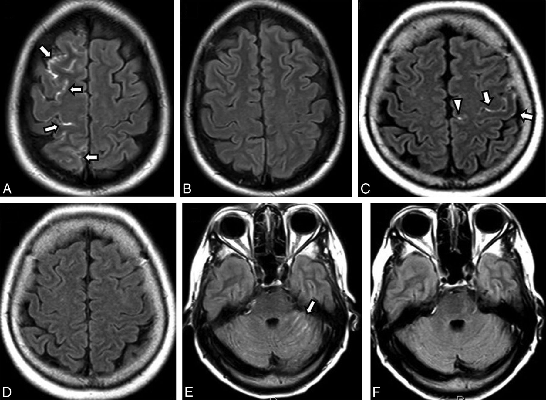
  • Fig 2.
    • Download figure
    • Open in new tab
    • Download powerpoint
    Fig 2.

    A, Right internal carotid artery angiographic approach for coiling of a right ophthalmic ICA aneurysm (black arrowhead) in a 71-year-old woman using 7.78 mL/kg of iodinated contrast volume and a procedural time of 4 hours. Note the presence of fetal origin of the right posterior cerebral artery (black arrow). B, Gadolinium-enhanced FLAIR performed at 12 hours after endovascular coiling shows cortical and sulcal hyperintensity along the right middle cerebral artery territory (white arrows) and the right PCA territory (white arrowheads). C, Corresponding DWI shows no diffusion restriction. D, A follow-up unenhanced CT scan obtained on the same date shows no evidence of subarachnoid hemorrhage. E, Two-month follow-up Gd-FLAIR shows no residual abnormality.

Tables

  • Figures
    • View popup
    Table 1:

    Patient demographics and aneurysms

    Patient data
        No.58
        Age (mean) (range) (yr)57.17 ± 10.94 (33–78)
        Sex (No.) (%)Female, 40 (69%)
        Endovascular coiling aneurysms (No.)62
    Target aneurysm locations (N = 62)
        AComA (No.) (%)10 (16.13)
        Right ICA (No.) (%)12 (19.35)
        Left ICA (No.) (%)8 (12.9)
        Right PcomA (No.) (%)4 (6.45)
        Right MCA (No.) (%)7 (11.29)
        Left MCA (No.) (%)2 (3.22)
        BA (No.) (%)17 (27.42)
        Left VA (No.) (%)1 (1.61)
        Left PCA (No.) (%)1 (1.61)
    Endovascular coiling techniques (N = 62)
        Routine coiling (No.) (%)23 (37.1%)
        Balloon-assisted coiling (No.) (%)29 (46.77%)
        Stent-assisted coiling (No.) (%)6 (9.68%)
        Pipeline flow diverter and coiling (No.) (%)3 (4.8%)
        Balloon- and stent-assisted coiling (No.) (%)1 (1.61%)
        No. of coils (median) (range)5 (1–32)
    • Note:—AcomA indicates anterior communicating artery; PcomA, posterior communicating artery; BA, basilar artery.

    • View popup
    Table 2:

    Comparison between findings negative and positive for CSHF

    FactorsFindings Negative for CSHF (n = 30)Findings Positive for CSHF (n = 32)Total (N = 62)P Value
    Contrast volume (mean) (mL)453.33 ± 156.98431.25 ± 161.52441.93 ± 158.42.588
    Contrast volume per weight (mean) (mL/kg)6.53 ± 2.396.15 ± 2.566.33 ± 2.46.556
    Duration of endovascular coiling procedure (median) (IQR) (hr)1.39 (0.95–2.20)2.21 (1.63–3.26)1.89 (1.26–2.66).001
    Time from coiling to MRI (median) (IQR) (hr)16.16 (11.99–22.51)16.08 (10.03–22.61)16.16 (11.56–22.125).871
    No. of angiographic runs (median) (IQR)13 (10–17)18 (12–22)14 (12–20).019
    DWI grading (No.) (%)
        091322 (35.5%).68
        I131225 (40.3%)
        II8715 (20.2%)
PreviousNext
Back to top

In this issue

American Journal of Neuroradiology: 39 (4)
American Journal of Neuroradiology
Vol. 39, Issue 4
1 Apr 2018
  • Table of Contents
  • Index by author
  • Complete Issue (PDF)
Advertisement
Print
Download PDF
Email Article

Thank you for your interest in spreading the word on American Journal of Neuroradiology.

NOTE: We only request your email address so that the person you are recommending the page to knows that you wanted them to see it, and that it is not junk mail. We do not capture any email address.

Enter multiple addresses on separate lines or separate them with commas.
HARMless: Transient Cortical and Sulcal Hyperintensity on Gadolinium-Enhanced FLAIR after Elective Endovascular Coiling of Intracranial Aneurysms
(Your Name) has sent you a message from American Journal of Neuroradiology
(Your Name) thought you would like to see the American Journal of Neuroradiology web site.
CAPTCHA
This question is for testing whether or not you are a human visitor and to prevent automated spam submissions.
Cite this article
S. Suthiphosuwan, C.C.-T. Hsu, A. Bharatha
HARMless: Transient Cortical and Sulcal Hyperintensity on Gadolinium-Enhanced FLAIR after Elective Endovascular Coiling of Intracranial Aneurysms
American Journal of Neuroradiology Apr 2018, 39 (4) 720-726; DOI: 10.3174/ajnr.A5561

Citation Manager Formats

  • BibTeX
  • Bookends
  • EasyBib
  • EndNote (tagged)
  • EndNote 8 (xml)
  • Medlars
  • Mendeley
  • Papers
  • RefWorks Tagged
  • Ref Manager
  • RIS
  • Zotero
0 Responses
Respond to this article
Share
Bookmark this article
HARMless: Transient Cortical and Sulcal Hyperintensity on Gadolinium-Enhanced FLAIR after Elective Endovascular Coiling of Intracranial Aneurysms
S. Suthiphosuwan, C.C.-T. Hsu, A. Bharatha
American Journal of Neuroradiology Apr 2018, 39 (4) 720-726; DOI: 10.3174/ajnr.A5561
del.icio.us logo Twitter logo Facebook logo Mendeley logo
  • Tweet Widget
  • Facebook Like
  • Google Plus One
Purchase

Jump to section

  • Article
    • Abstract
    • ABBREVIATIONS:
    • Materials and Methods
    • Results
    • Discussion
    • Conclusions
    • References
  • Figures & Data
  • Info & Metrics
  • Responses
  • References
  • PDF

Related Articles

  • No related articles found.
  • PubMed
  • Google Scholar

Cited By...

  • No citing articles found.
  • Crossref (7)
  • Google Scholar

This article has been cited by the following articles in journals that are participating in Crossref Cited-by Linking.

  • No association between cortical lesions and leptomeningeal enhancement on 7-Tesla MRI in multiple sclerosis
    Mehrnaz Ighani, Samuel Jonas, Izlem Izbudak, Seongjin Choi, Alfonso Lema-Dopico, Jun Hua, Erin E O’Connor, Daniel M Harrison
    Multiple Sclerosis Journal 2020 26 2
  • CSF enhancement on post-contrast fluid-attenuated inversion recovery images; a systematic review
    Whitney M. Freeze, Merel van der Thiel, Jeroen de Bresser, Catharina J.M. Klijn, Ellis S. van Etten, Jacobus F.A. Jansen, Louise van der Weerd, Heidi I.L. Jacobs, Walter H. Backes, Susanne J. van Veluw
    NeuroImage: Clinical 2020 28
  • Leptomeningeal Enhancement Is Associated with Transient Neurologic Deficits after Flow Diversion of Intracranial Aneurysms
    Yiping Li, Chadd Funk, Demi Dawkins, Dustin Simpson, John-Paul J. Yu, Melanie Boly, Azam Ahmed
    World Neurosurgery 2018 120
  • Difference in imaging biomarkers between transient and permanent neurological deficits after endovascular treatment of cerebrovascular aneurysms
    Yiping Li, Jason Kim, Dustin Simpson, Beverly Aagaard-Kienitz, David Niemann, Ignatius N. Esene, Azam Ahmed
    Journal of Neurosurgery 2021 134 6
  • Contrast enhanced FLAIR versus contrast enhanced T1W images in evaluation of intraparenchymal brain lesions
    Mohamed Ali EL-Adalany, Maha Mohamed Zaki Bilal, Yomna Abdulwahab Mohmamed Rihan, Dina EL-Metwally
    Egyptian Journal of Radiology and Nuclear Medicine 2022 53 1
  • Contrast-enhanced encephalopathy and massive cerebral edema after endovascular coiling of cerebral aneurysm. A case report
    Zhou Jia-hao, Seidu A. Richard, Deng Yin-sheng, Jiang Ming, Zhuang Yan
    Veins and Lymphatics 2021 10 1
  • Spectrum and Mimics of Brain Surface Enhancement on Postcontrast FLAIR Images
    S.N. Rogers, J.L. Becker
    Neurographics 2023 13 2

More in this TOC Section

ADULT BRAIN

  • Diagnostic Neuroradiology of Monoclonal Antibodies
  • Clinical Outcomes After Chiari I Decompression
  • Segmentation of Brain Metastases with BLAST
Show more ADULT BRAIN

INTERVENTIONAL

  • SAVE vs. Solumbra Techniques for Thrombectomy
  • Contrast-Induced Encephalopathy after NeuroIR
  • CT Perfusion&Reperfusion in Acute Ischemic Stroke
Show more INTERVENTIONAL

Similar Articles

Advertisement

Indexed Content

  • Current Issue
  • Accepted Manuscripts
  • Article Preview
  • Past Issues
  • Editorials
  • Editor's Choice
  • Fellows' Journal Club
  • Letters to the Editor
  • Video Articles

Cases

  • Case Collection
  • Archive - Case of the Week
  • Archive - Case of the Month
  • Archive - Classic Case

Special Collections

  • AJNR Awards
  • ASNR Foundation Special Collection
  • Most Impactful AJNR Articles
  • Photon-Counting CT
  • Spinal CSF Leak Articles (Jan 2020-June 2024)

More from AJNR

  • Trainee Corner
  • Imaging Protocols
  • MRI Safety Corner

Multimedia

  • AJNR Podcasts
  • AJNR Scantastics

Resources

  • Turnaround Time
  • Submit a Manuscript
  • Submit a Video Article
  • Submit an eLetter to the Editor/Response
  • Manuscript Submission Guidelines
  • Statistical Tips
  • Fast Publishing of Accepted Manuscripts
  • Graphical Abstract Preparation
  • Imaging Protocol Submission
  • Evidence-Based Medicine Level Guide
  • Publishing Checklists
  • Author Policies
  • Become a Reviewer/Academy of Reviewers
  • News and Updates

About Us

  • About AJNR
  • Editorial Board
  • Editorial Board Alumni
  • Alerts
  • Permissions
  • Not an AJNR Subscriber? Join Now
  • Advertise with Us
  • Librarian Resources
  • Feedback
  • Terms and Conditions
  • AJNR Editorial Board Alumni

American Society of Neuroradiology

  • Not an ASNR Member? Join Now

© 2025 by the American Society of Neuroradiology All rights, including for text and data mining, AI training, and similar technologies, are reserved.
Print ISSN: 0195-6108 Online ISSN: 1936-959X

Powered by HighWire