
Article Figures & Data
Figures
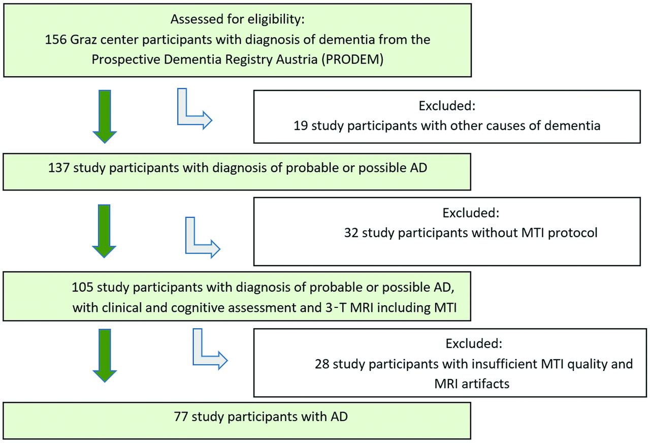
- FIG 1.
Flowchart shows the recruitment of the study participants with AD.
Tables
Study Participant Characteristic AD (n = 77) Healthy Control Participants (n = 77) P Valueb No. female (%) 47 (61) 44 (57) .62 Age, years,a 72 (8) 72 (8) .98 Age range, years 51–87 51–87 MMSEa 22.03 (3.72) 27.57 (1.75) <.001 Global cortex volume, cm3a 329.63 (14.53) 394.04 (41.25) <.001 AD-signature regions volume, cm3a 54.81 (10.15) 70.28 (8.86) <.001 Global NAWM volume, cm3a 305.77 (63.71) 334.70 (52.52) .01 WM hyperintensities volume, cm3a 16.19 (17.69) 11.64 (18.01) .02 Fazekas grade 2 or 3, no. (%) 49 (63.7) 31 (40.3) Global cortex MTRsa 0.295 (0.016) 0.309 (0.008) <.001 AD-signature regions MTRsa 0.297 (0.018) 0.309 (0.009) <.001 Global NAWM MTRsa 0.384 (0.009) 0.388 (0.009) .003 WM hyperintensities MTRsa 0.322 (0.028) 0.350 (0.016) <.001 - Table 2:
Logistic regression analysis—lower MTRs relate to AD independent of normalized regional brain volume and white matter damage
MTR Region OR 95% CI P Valuea Global cortex 0.47 0.22–0.97 .04 AD-signature regions 0.31 0.14–0.67 .003 Global NAWM 0.59 0.39–0.88 .01 WM hyperintensities 0.18 0.09–0.33 <.001 ↵a Corrected for age, sex, years of education, normalized regional volume, and Fazekas score.
- Table 3:
Linear regression analysis in AD—cortical MTRs relate to poorer language function in patients with AD
MTR Region MMSE, n = 77 Language Function (CERAD Test: Boston Naming Test), n = 77 Constructional Praxis (CERAD Test: Figure Copying), n = 73 β 95% CI P Valuea β 95% CI P Valuea β 95% CI P Valuea Global cortex 0.23 −0.07–1.96 .06 0.31 0.19–1.95 .02 0.22 −0.09–1.24 .09 AD-signature regions 0.14 −0.50–1.52 .32 0.28 −0.02–1.74 .05 0.29 −0.01–1.32 .05 Global NAWM 0.17 −0.24–1.67 .14 0.11 −0.05–1.23 .40 0.23 −0.05–1.23 .07 WM hyperintensities 0.01 −0.78–0.88 .91 -0.01 −0.81–0.67 .89 0.01 −0.53–0.59 .92 Note:—β indicates standardized regression coefficient.
↵a Corrected for age, sex, years of education, normalized regional volume, and Fazekas score.


Jump to section
Related Articles
Cited By...
- No citing articles found.