Advertisement

Main menu

  • Home
  • Content
    • Current Issue
    • Accepted Manuscripts
    • Article Preview
    • Past Issue Archive
    • Video Articles
    • AJNR Case Collection
    • Case of the Week Archive
    • Case of the Month Archive
    • Classic Case Archive
  • Special Collections
    • AJNR Awards
    • ASNR Foundation Special Collection
    • Most Impactful AJNR Articles
    • Photon-Counting CT
    • Spinal CSF Leak Articles (Jan 2020-June 2024)
  • Multimedia
    • AJNR Podcasts
    • AJNR SCANtastic
    • Trainee Corner
    • MRI Safety Corner
    • Imaging Protocols
  • For Authors
    • Submit a Manuscript
    • Submit a Video Article
    • Submit an eLetter to the Editor/Response
    • Manuscript Submission Guidelines
    • Statistical Tips
    • Fast Publishing of Accepted Manuscripts
    • Graphical Abstract Preparation
    • Imaging Protocol Submission
    • Author Policies
  • About Us
    • About AJNR
    • Editorial Board
    • Editorial Board Alumni
  • More
    • Become a Reviewer/Academy of Reviewers
    • Subscribers
    • Permissions
    • Alerts
    • Feedback
    • Advertisers
    • ASNR Home

User menu

  • Alerts
  • Log in

Search

  • Advanced search
American Journal of Neuroradiology

American Journal of Neuroradiology

ASHNR American Society of Functional Neuroradiology ASHNR American Society of Pediatric Neuroradiology ASSR
  • Alerts
  • Log in

Advanced Search

  • Home
  • Content
    • Current Issue
    • Accepted Manuscripts
    • Article Preview
    • Past Issue Archive
    • Video Articles
    • AJNR Case Collection
    • Case of the Week Archive
    • Case of the Month Archive
    • Classic Case Archive
  • Special Collections
    • AJNR Awards
    • ASNR Foundation Special Collection
    • Most Impactful AJNR Articles
    • Photon-Counting CT
    • Spinal CSF Leak Articles (Jan 2020-June 2024)
  • Multimedia
    • AJNR Podcasts
    • AJNR SCANtastic
    • Trainee Corner
    • MRI Safety Corner
    • Imaging Protocols
  • For Authors
    • Submit a Manuscript
    • Submit a Video Article
    • Submit an eLetter to the Editor/Response
    • Manuscript Submission Guidelines
    • Statistical Tips
    • Fast Publishing of Accepted Manuscripts
    • Graphical Abstract Preparation
    • Imaging Protocol Submission
    • Author Policies
  • About Us
    • About AJNR
    • Editorial Board
    • Editorial Board Alumni
  • More
    • Become a Reviewer/Academy of Reviewers
    • Subscribers
    • Permissions
    • Alerts
    • Feedback
    • Advertisers
    • ASNR Home
  • Follow AJNR on Twitter
  • Visit AJNR on Facebook
  • Follow AJNR on Instagram
  • Join AJNR on LinkedIn
  • RSS Feeds

AJNR is seeking candidates for the AJNR Podcast Editor. Read the position description.

Research ArticleFUNCTIONAL

White Matter Microstructural Abnormalities in Type 2 Diabetes Mellitus: A Diffusional Kurtosis Imaging Analysis

Y. Xie, Y. Zhang, W. Qin, S. Lu, C. Ni and Q. Zhang
American Journal of Neuroradiology March 2017, 38 (3) 617-625; DOI: https://doi.org/10.3174/ajnr.A5042
Y. Xie
aFrom the Department of Radiology and Tianjin Key Laboratory of Functional Imaging (Y.X., Y.Z., W.Q., Q.Z.), Tianjin Medical University General Hospital, Tianjin, China
  • Find this author on Google Scholar
  • Find this author on PubMed
  • Search for this author on this site
  • ORCID record for Y. Xie
Y. Zhang
aFrom the Department of Radiology and Tianjin Key Laboratory of Functional Imaging (Y.X., Y.Z., W.Q., Q.Z.), Tianjin Medical University General Hospital, Tianjin, China
  • Find this author on Google Scholar
  • Find this author on PubMed
  • Search for this author on this site
  • ORCID record for Y. Zhang
W. Qin
aFrom the Department of Radiology and Tianjin Key Laboratory of Functional Imaging (Y.X., Y.Z., W.Q., Q.Z.), Tianjin Medical University General Hospital, Tianjin, China
  • Find this author on Google Scholar
  • Find this author on PubMed
  • Search for this author on this site
  • ORCID record for W. Qin
S. Lu
bDepartments of Radiology (S.L.)
  • Find this author on Google Scholar
  • Find this author on PubMed
  • Search for this author on this site
  • ORCID record for S. Lu
C. Ni
cCardiology (C.N.), Tianjin Medical University Metabolic Diseases Hospital, Tianjin, China.
  • Find this author on Google Scholar
  • Find this author on PubMed
  • Search for this author on this site
  • ORCID record for C. Ni
Q. Zhang
aFrom the Department of Radiology and Tianjin Key Laboratory of Functional Imaging (Y.X., Y.Z., W.Q., Q.Z.), Tianjin Medical University General Hospital, Tianjin, China
  • Find this author on Google Scholar
  • Find this author on PubMed
  • Search for this author on this site
  • ORCID record for Q. Zhang
  • Article
  • Figures & Data
  • Supplemental
  • Info & Metrics
  • Responses
  • References
  • PDF
Loading

Article Figures & Data

Figures

  • Tables
  • Fig 1.
    • Download figure
    • Open in new tab
    • Download powerpoint
    Fig 1.

    WM regions showed significant differences in DKI metrics between patients with T2DM and controls. The regions with decreased FA (upper row), increased MD (middle row), and decreased MK (lower row) in patients with T2DM were overlapped on the FA template. Color bars represent the t values of intergroup comparisons.
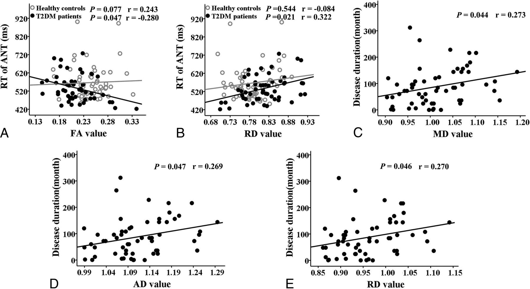
  • Fig 2.
    • Download figure
    • Open in new tab
    • Download powerpoint
    Fig 2.

    Correlations between DKI parameters and clinical/cognitive variables in the patients with T2DM and controls. A, Correlation between decreased FA in the rPF_WM and RT of ANT. B, Correlation between increased RD in rPF_WM and RT of ANT. C, Correlation between increased MD in the superior rPF_WM and disease duration. D, Correlation between increased AD in the superior rPF_WM and disease duration. E, Correlation between increased RD in the superior rPF_WM and disease duration.

  • Fig 3.
    • Download figure
    • Open in new tab
    • Download powerpoint
    Fig 3.

    WM regions that showed significant differences in DKI metrics (red) and DTI metrics (blue) between patients with T2DM and controls were overlapped on the FA template. Green represents the overlapped region of the results of the DKI and the DTI between patients with T2DM and controls.

Tables

  • Figures
    • View popup
    Table 1:

    Demographic and clinical informationa

    T2DM (n = 58)Healthy Controls (n = 58)StatisticsP
    Demographics
        Age (yr)56.09 ± 8.1654.66 ± 7.03t = 1.012.314
        Sex (M/F)34:2435:23χ2 = 0.036.850
        Education level (yr)11.72 ± 3.3111.07 ± 2.64t = 1.178.241
    Clinical information
        Disease duration (mo)91.25 ± 69.81–––
        BMI (kg/m2)25.57 ± 2.1924.64 ± 2.99t = 1.915.058
        Systolic BP (mm Hg)130 (110–170)122.5 (100–170)z = −1.713.087
        Diastolic BP (mm Hg)80 (60–100)80 (60–100)z = −1.015.310
        FBG (mmol/L)8.06 ± 2.815.13 ± 0.65t = 7.717<.001
        HbA1c (%)8.35 ± 2.105.56 ± 0.33t = 10.020<.001
        HbA1c (mmol/mol)67.76 ± 22.8737.31 ± 3.62t = 10.013<.001
        Total cholesterol (mmol/L)5.06 ± 1.355.39 ± 0.91t = −1.541.126
        Triglycerides (mmol/L)1.66 (0.49–8.03)1.24 (0.46–7.35)z = −1.698.090
        HDL (mmol/L)1.15 ± 0.271.27 ± 0.31t = −2.167.032
        LDL (mmol/L)3.17 ± 0.933.47 ± 0.85t = −1.799.075
    • Note:—FBG indicates fasting blood glucose; HDL, high-density lipoprotein; LDL, low-density lipoprotein; BP, blood pressure; BMI, body mass index.

    • ↵a Data distributed normally or non-normally are presented as mean ± SD or median (range).

    • View popup
    Table 2:

    Cognitive informationa

    T2DM (n = 58)Healthy Controls (n = 58)StatisticsP
    MMSE score29.21 ± 0.8929.50 ± 0.94t = −1.720.088
    RT of ANT (ms)536.53 ± 71.31566.51 ± 92.09t = −1.911.059
    ACC of ANT (%)98.65 ± 1.8397.89 ± 4.49t = 1.157.250
    RPEP (%)0.07 ± 0.020.08 ± 0.02t = −1.908.059
    SAS32.19 ± 6.1630.71 ± 5.24t = 1.397.165
    SDS34.09 ± 7.7931.64 ± 7.07t = 1.772.079
    Long-term memory10.75 ± 2.6511.04 ± 2.85t = −0.555.580
    Short-term memory47.14 ± 9.2548.69 ± 9.29t = −0.879.381
    Forward Digit Span8.25 ± 1.508.18 ± 1.26t = 0.258.797
    Backward Digit Span5.07 ± 1.455.13 ± 1.07t = −0.230.818
    TMT-A (s)61.65 ± 25.5458.15 ± 27.60t = 0.686.494
    • Note:—ACC of ANT indicates the accuracy rate of Attention Network Test; RPEP, the percentage of the preservative response error; SAS, Self-Rating Anxiety Scale; SDS, Self-Rating Depressive Scale; TMT-A, Trail Making Test A; MMSE, Mini-Mental State Examination.

    • ↵a Data are presented as mean ± SD.

    • View popup
    Table 3:

    Brain regions showing significant voxel-based intergroup differences in DKI metrics

    Structure NameMetricsCluster Voxel No.Peak t ValueMNI Coordinates
    XYZ
    Splenium of CCFA184−4.679−2−348
    rPF_WMFA78−4.483302620
    Superior rPF_WMMD2045.094103636
    Middle rPF_WMMD5215.077145210
    Inferior rPF_WMMD903.9003842−2
    lPF_WMMD804.372−463226
    rST_WMMD1894.608340−22
    Splenium of CCMD753.882−2−388
    lECMD2615.068−3212−2
    PonsMD1334.487−6−28−42
    Splenium of CCMK172−4.4270−3214
    PonsMK150−4.659−6−28−42
    • Note:—lPF_WM indicates left prefrontal white matter; MNI, Montreal Neurological Institute; rST_WM, right superior temporal white matter.

    • View popup
    Table 4:

    Intergroup differences of the AD and RD values that contributed to the altered FA valuesa

    T2DM (n = 58)Healthy Controls (n = 58)FP
    rPF_WM
        AD, ×10−5 mm2/s111.10 ± 4.83111.79 ± 4.630.967.328
        RD, ×10−5 mm2/s82.06 ± 5.4878.84 ± 4.7112.095.001
    Splenium of CC
        AD, ×10−5 mm2/s288.14 ± 26.33270.95 ± 33.148.490.004
        RD, ×10−5 mm2/s179.24 ± 36.37151.55 ± 41.7713.321<.001
    • ↵a Data are presented as mean ± SD. The intergroup comparisons were performed with ANCOVA adjusted for age, sex, and years of education.

    • View popup
    Table 5:

    Intergroup differences of the AD and RD values that contributed to the altered MD valuesa

    T2DM (n = 58)Healthy Controls (n = 58)FP
    Superior rPF_WM
        AD, ×10−5 mm2/s111.06 ± 6.52105.23 ± 4.2531.431<.001
        RD, ×10−5 mm2/s93.28 ± 6.4187.75 ± 4.4928.488<.001
    Middle rPF_WM
        AD, ×10−5 mm2/s113.59 ± 6.04108.52 ± 3.4831.472<.001
        RD, ×10−5 mm2/s90.12 ± 6.3985.27 ± 3.5327.982<.001
    Inferior rPF_WM
        AD, ×10−5 mm2/s116.85 ± 5.98112.19 ± 5.5318.548<.001
        RD, ×10−5 mm2/s84.53 ± 5.9581.24 ± 4.3810.752.001
    lPF_WM
        AD, ×10−5 mm2/s121.75 ± 10.54114.50 ± 6.6219.725<.001
        RD, ×10−5 mm2/s107.47 ± 11.4098.84 ± 7.4423.252<.001
    rST_WM
        AD, ×10−5 mm2/s127.10 ± 10.59119.03 ± 6.3822.413<.001
        RD, ×10−5 mm2/s102.70 ± 10.2594.47 ± 5.8525.394<.001
    Splenium of CC
        AD, ×10−5 mm2/s269.21 ± 20.44255.20 ± 24.279.865.002
        RD, ×10−5 mm2/s152.25 ± 32.11127.36 ± 32.8415.805<.001
    lEC
        AD, ×10−5 mm2/s122.54 ± 7.42117.14 ± 4.8421.495<.001
        RD, ×10−5 mm2/s92.85 ± 8.7086.36 ± 6.1422.007<.001
    Pons
        AD, ×10−5 mm2/s222.11 ± 27.07201.58 ± 26.1518.416<.001
        RD, ×10−5 mm2/s177.25 ± 27.60157.84 ± 24.3817.237<.001
    • Note:—lPF_WM indicates left prefrontal white matter; rST_WM, right superior temporal white matter.

    • ↵a Data are presented as mean ± SD. The intergroup comparisons were performed with ANCOVA adjusted for age, sex, and years of education.

    • View popup
    Table 6:

    Intergroup differences of the AK and RK values that contributed to the altered MK valuesa

    T2DM (n = 58)Healthy Controls (n = 58)FP
    Splenium of CC
        AK, ×10−5 mm2/s46.10 ± 2.3947.90 ± 3.1611.249.001
        RK, ×10−5 mm2/s112.35 ± 22.24131.37 ± 26.6216.883<.001
    Pons
        AK, ×10−5 mm2/s66.02 ± 6.1971.75 ± 8.0918.289<.001
        RK, ×10−5 mm2/s83.59 ± 8.9890.75 ± 8.8417.912<.001
    • ↵a Data are presented as mean ± SD. The intergroup comparisons were performed with ANCOVA adjusted for age, sex, and years of education.

PreviousNext
Back to top

In this issue

American Journal of Neuroradiology: 38 (3)
American Journal of Neuroradiology
Vol. 38, Issue 3
1 Mar 2017
  • Table of Contents
  • Index by author
  • Complete Issue (PDF)
Advertisement
Print
Download PDF
Email Article

Thank you for your interest in spreading the word on American Journal of Neuroradiology.

NOTE: We only request your email address so that the person you are recommending the page to knows that you wanted them to see it, and that it is not junk mail. We do not capture any email address.

Enter multiple addresses on separate lines or separate them with commas.
White Matter Microstructural Abnormalities in Type 2 Diabetes Mellitus: A Diffusional Kurtosis Imaging Analysis
(Your Name) has sent you a message from American Journal of Neuroradiology
(Your Name) thought you would like to see the American Journal of Neuroradiology web site.
CAPTCHA
This question is for testing whether or not you are a human visitor and to prevent automated spam submissions.
Cite this article
Y. Xie, Y. Zhang, W. Qin, S. Lu, C. Ni, Q. Zhang
White Matter Microstructural Abnormalities in Type 2 Diabetes Mellitus: A Diffusional Kurtosis Imaging Analysis
American Journal of Neuroradiology Mar 2017, 38 (3) 617-625; DOI: 10.3174/ajnr.A5042

Citation Manager Formats

  • BibTeX
  • Bookends
  • EasyBib
  • EndNote (tagged)
  • EndNote 8 (xml)
  • Medlars
  • Mendeley
  • Papers
  • RefWorks Tagged
  • Ref Manager
  • RIS
  • Zotero
0 Responses
Respond to this article
Share
Bookmark this article
White Matter Microstructural Abnormalities in Type 2 Diabetes Mellitus: A Diffusional Kurtosis Imaging Analysis
Y. Xie, Y. Zhang, W. Qin, S. Lu, C. Ni, Q. Zhang
American Journal of Neuroradiology Mar 2017, 38 (3) 617-625; DOI: 10.3174/ajnr.A5042
del.icio.us logo Twitter logo Facebook logo Mendeley logo
  • Tweet Widget
  • Facebook Like
  • Google Plus One
Purchase

Jump to section

  • Article
    • Abstract
    • ABBREVIATIONS:
    • Materials and Methods
    • Results
    • Discussion
    • Conclusions
    • Footnotes
    • References
  • Figures & Data
  • Supplemental
  • Info & Metrics
  • Responses
  • References
  • PDF

Related Articles

  • PubMed
  • Google Scholar

Cited By...

  • White Matter Microstructural Alteration in Type 2 Diabetes: A Combined UK Biobank Study of Diffusion Tensor Imaging and Neurite Orientation Dispersion and Density Imaging
  • Crossref (25)
  • Google Scholar

This article has been cited by the following articles in journals that are participating in Crossref Cited-by Linking.

  • Traumatic Brain Injury: Imaging Patterns and Complications
    Andrew D. Schweitzer, Sumit N. Niogi, Christopher J. Whitlow, A. John Tsiouris
    RadioGraphics 2019 39 6
  • Brain microstructural abnormalities in type 2 diabetes mellitus: A systematic review of diffusion tensor imaging studies
    Hossein Sanjari Moghaddam, Farzaneh Ghazi Sherbaf, Mohammad Hadi Aarabi
    Frontiers in Neuroendocrinology 2019 55
  • Glucose disturbances, cognitive deficits and white matter abnormalities in first-episode drug-naive schizophrenia
    Xiangyang Zhang, Mi Yang, Xiangdong Du, Wei Liao, Dachun Chen, Fengmei Fan, Meihong Xiu, Qiufang Jia, Yuping Ning, Xingbing Huang, Fengchun Wu, Jair C. Soares, Bo Cao, Li Wang, Huafu Chen
    Molecular Psychiatry 2020 25 12
  • Abnormal brain white matter in patients with right trigeminal neuralgia: a diffusion tensor imaging study
    Junpeng Liu, Jiajia Zhu, Fei Yuan, Xuejun Zhang, Quan Zhang
    The Journal of Headache and Pain 2018 19 1
  • Brain microstructural alterations in type 2 diabetes: diffusion kurtosis imaging provides added value to diffusion tensor imaging
    Ying Xiong, Yi Sui, Shun Zhang, Xiaohong Joe Zhou, Shaolin Yang, Yang Fan, Qiang Zhang, Wenzhen Zhu
    European Radiology 2019 29 4
  • Altered brain structural topological properties in type 2 diabetes mellitus patients without complications
    Yang Zhang, Yujuan Cao, Yingjie Xie, Linlin Liu, Wen Qin, Shan Lu, Quan Zhang
    Journal of Diabetes 2019 11 2
  • Investigating Brain Microstructural Alterations in Type 1 and Type 2 Diabetes Using Diffusion Tensor Imaging: A Systematic Review
    Abdulmajeed Alotaibi, Christopher Tench, Rebecca Stevenson, Ghadah Felmban, Amjad Altokhis, Ali Aldhebaib, Rob A. Dineen, Cris S. Constantinescu
    Brain Sciences 2021 11 2
  • Elevated Insulin and Insulin Resistance are Associated with Altered Myelin in Cognitively Unimpaired Middle‐Aged Adults
    J. Patrick O’Grady, Douglas C. Dean, Kao Lee Yang, Cristybelle‐Marie Canda, Siobhan M. Hoscheidt, Erika J. Starks, Andrew Merluzzi, Samuel Hurley, Nancy J. Davenport, Ozioma C. Okonkwo, Rozalyn M. Anderson, Sanjay Asthana, Sterling C. Johnson, Andrew L. Alexander, Barbara B. Bendlin
    Obesity 2019 27 9
  • Abnormalities of Brain White Matter in Type 2 Diabetes Mellitus: A Meta-Analysis of Diffusion Tensor Imaging
    Li Huang, Qingqing Zhang, Tong Tang, Minguang Yang, Cong Chen, Jing Tao, Shengxiang Liang
    Frontiers in Aging Neuroscience 2021 13
  • Alterations of Brain Structural Network Connectivity in Type 2 Diabetes Mellitus Patients With Mild Cognitive Impairment
    Chang Li, Jingna Zhang, Mingguo Qiu, Kaijun Liu, Yang Li, Zhiwei Zuo, Xuntao Yin, Yuqi Lai, Jingqin Fang, Haipeng Tong, Yu Guo, Jian Wang, Xiao Chen, Kunlin Xiong
    Frontiers in Aging Neuroscience 2021 12

More in this TOC Section

FUNCTIONAL

  • Kurtosis and Epileptogenic Tubers: A Pilot Study
  • Glutaric Aciduria Type 1: DK vs. Conventional MRI
  • Predicting Outcomes in Tuberous Sclerosis Epilepsy
Show more FUNCTIONAL

ADULT BRAIN

  • Diagnostic Neuroradiology of Monoclonal Antibodies
  • Cerebral ADC Changes in Fabry Disease
  • ML for Glioma Molecular Subtype Prediction
Show more ADULT BRAIN

Similar Articles

Advertisement

Indexed Content

  • Current Issue
  • Accepted Manuscripts
  • Article Preview
  • Past Issues
  • Editorials
  • Editor's Choice
  • Fellows' Journal Club
  • Letters to the Editor
  • Video Articles

Cases

  • Case Collection
  • Archive - Case of the Week
  • Archive - Case of the Month
  • Archive - Classic Case

Special Collections

  • AJNR Awards
  • ASNR Foundation Special Collection
  • Most Impactful AJNR Articles
  • Photon-Counting CT
  • Spinal CSF Leak Articles (Jan 2020-June 2024)

More from AJNR

  • Trainee Corner
  • Imaging Protocols
  • MRI Safety Corner

Multimedia

  • AJNR Podcasts
  • AJNR Scantastics

Resources

  • Turnaround Time
  • Submit a Manuscript
  • Submit a Video Article
  • Submit an eLetter to the Editor/Response
  • Manuscript Submission Guidelines
  • Statistical Tips
  • Fast Publishing of Accepted Manuscripts
  • Graphical Abstract Preparation
  • Imaging Protocol Submission
  • Evidence-Based Medicine Level Guide
  • Publishing Checklists
  • Author Policies
  • Become a Reviewer/Academy of Reviewers
  • News and Updates

About Us

  • About AJNR
  • Editorial Board
  • Editorial Board Alumni
  • Alerts
  • Permissions
  • Not an AJNR Subscriber? Join Now
  • Advertise with Us
  • Librarian Resources
  • Feedback
  • Terms and Conditions
  • AJNR Editorial Board Alumni

American Society of Neuroradiology

  • Not an ASNR Member? Join Now

© 2025 by the American Society of Neuroradiology All rights, including for text and data mining, AI training, and similar technologies, are reserved.
Print ISSN: 0195-6108 Online ISSN: 1936-959X

Powered by HighWire